

Reference 04042 · Anatomical model
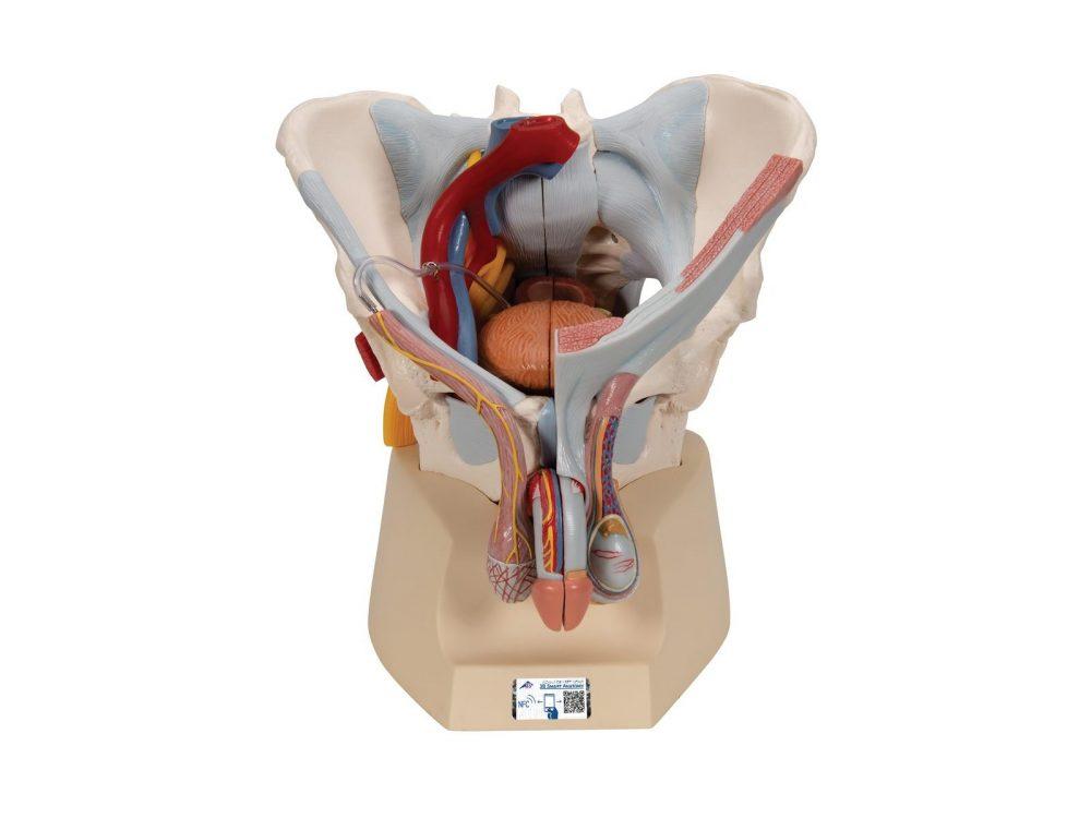
Male pelvis model with ligaments, vessels, nerves, pelvic floor and organs - 7 Parts
- Anatomical model
- Orthopaedic surgery
- Main product
- Medicine
This 7 part model of the male pelvis shows in accurate detail how the bones, ligaments, vessels and nerves as well as the pelvic floor muscles and the external sex organs are connected to each other. It shows the whole pelvis, through which a median section has been placed.
Price on request
Available in Spain, Portugal and Andorra · delivery time given in the proposal
Downloadable data sheet — features, specifications and delivery time, in PDF.
Need a technical report? Ask us for it and we'll send it within 24 h →
Description
What this equipment is and what it's designed for.
This 7 part model of the male pelvis shows in accurate detail how the bones, ligaments, vessels and nerves as well as the pelvic floor muscles and the external sex organs are connected to each other. It shows the whole pelvis, through which a median section has been placed. The right side of the external anal sphincter, the M. ischiocavernosus, the M. transversus perinei profundus and superficialis and the M. bulbospongiosus can be removed together.
The rectum, bladder, prostate and penis can also be removed, and split into two halves at median level – partially connected. The bone structures are connected with magnets and can therefore be easily taken apart. The skin and the fascia of the penis have been partly removed so that the vessels and nerves are visible. Part of the skin in the area of the scrotum and the spermatic cord has removed too. The testicles and epididymis are also visible. On the left, the spermatic cord has been opened up in layers, and on the right the M. cremaster and fascia spermatica interna have been exposed.
The right half of the pelvis shows sections of the common, external and internal iliac arteries and the common and external iliac veins, demonstrating their positions in relation to each other. The Plexus sacralis, the N. ischiadicus, the N. pudendus, the N. dorsalis penis, the Nn. scrotales anteriores, the Nn. perineales and the Nn. annales inferiores as well as the Ductus deferens are shown. The model shows the following bones and ligaments: both hip bones, pubic symphisis, sacrum and coccyx as well as the fifth lumbar vertebra with intervertebral disc. A median section has been placed through the fifth lumbar vertebra, the sacrum and the coccyx, so that the pelvis can be split into two halves. This means that part of the cauda equina is also visible in the vertebral canal. The left half of the fifth lumbar vertebra can be removed.
The following ligament structures of the pelvis are shown:
- Lig. inguinale
- Lig. sacrotuberale
- Lig. sacrospinale
- Lig. sacroiliaca anteriora
- Lig. iliolumbale
- Lig. longitudinale anterius
- Lig. Supraspinale, Lig. sacroiliacum interosseum
- Lig. sacroiliacum posterius
- Lig. sacrococcygeum laterale
- Lig. sacrococcygeum posterius superficiale et profundum
- Membrana obturatoria
- Lig. lacunare
What you learn
Anatomical model modality. The competencies this equipment trains, in the language of the training plan.
- Identification of the bones and ligaments of the male pelvis
- Recognition of the pelvic floor musculature
- Location of the iliac vessels and their divisions
- Identification of the sacral plexus, the sciatic nerve and the pudendal nerve
- Description of the anatomy of the penis, the scrotum and the spermatic cord
- Assembly and disassembly of the removable structures
Cases and pathologies
The clinical scenarios that can be simulated with this equipment.
- Benign prostatic hyperplasia and its relationship with the bladder and the rectum
- Male pelvic floor dysfunction
- Pudendal nerve block
- Pathology of the scrotal contents: testis, epididymis and spermatic cord
- Urinary catheterisation and the course of the male urethra
Features and what's included
What the equipment does and what comes in the box, as described by the manufacturer.
Features
- Model of the male pelvis, dismantles into 7 parts
- Complete pelvic floor sectioned along the midline; the right portion of the external anal sphincter, the ischiocavernosus, the deep and superficial transverse perineal muscles and the bulbospongiosus are removed together
- Rectum, urinary bladder, prostate and penis removable and separable through their midsection
- Bony structure joined by magnets, separable into all its parts
- Penis with the fasciae and the skin partially removed, exposing vessels and nerves
- Scrotum and spermatic cord with the skin partially removed: testes and epididymides exposed; spermatic cord opened in layers on the left side and cremaster muscle and spermatic fascia exposed on the right
- Right half with the common, external and internal iliac arteries and the common and external iliac veins
- Sacral plexus, sciatic nerve, pudendal nerve, dorsal nerve of the penis, anterior scrotal nerves, perineal nerves and inferior anal nerves
- Bones: both hip bones, pubic symphysis, sacrum, coccyx and fifth lumbar vertebra; the left half of the vertebral body separates by means of a magnet and reveals a portion of the cauda equina
- Ligaments represented: inguinal, sacrotuberous, sacrospinous, anterior sacroiliac, iliolumbar, anterior longitudinal, supraspinous, interosseous sacroiliac, posterior sacroiliac, lateral sacrococcygeal, superficial and deep posterior sacrococcygeal, obturator membrane and lacunar ligament
Services for this equipment
Services matter: the equipment gets commissioned, the staff know how to use it and maintenance is settled before you start. Include them in your budget and avoid having to raise a second purchase.
- Preventive maintenance We'll let you know when it's due
Documentation
You can download it now or we'll send it on request.
Other related equipment
They share a specialty with this one. They train different things: none of them replaces it.
-
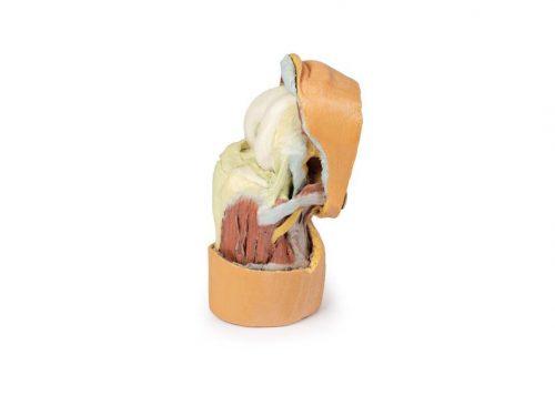 3D ANATOMY - Deep dissection of the knee joint, flexed
See details →
-
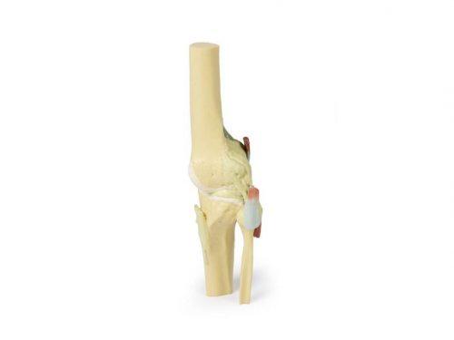 3D ANATOMY - Knee joint, extended
See details →
-
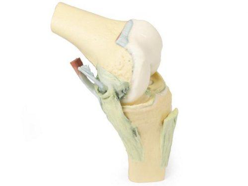 3D ANATOMY - Knee joint, flexed
See details →
-
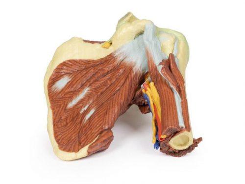 3D ANATOMY - Left shoulder, deep dissection of the joint, musculature, nerves and vessels
See details →
-
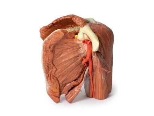 3D ANATOMY - Left shoulder, superficial muscles and axillary/brachial artery
See details →
-
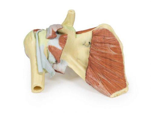 3D ANATOMY - Right shoulder, deep dissection of shoulder girdle, lateral clavicle and humerus
See details →
Is this equipment going into a new centre?
If you're equipping a classroom or a whole centre, starting from the shopping list works out expensive. Start from the needs assessment.