

Reference 02185 · Anatomical model
Functional knee joint
- Anatomical model
- Orthopaedic surgery
- Main product
- Medicine
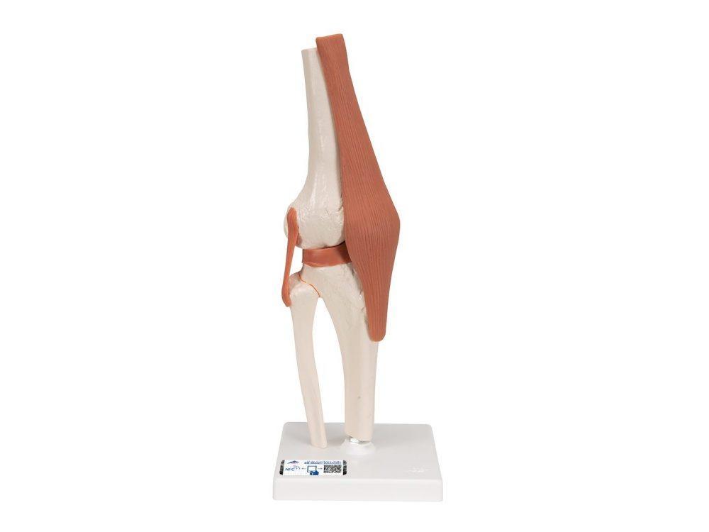
This high-quality, life-size knee joint model accurately demonstrates the structure and movement of the human knee joint. Designed for anatomical education and clinical demonstration, the model provides a clear understanding of knee biomechanics and joint function.
Price on request
Available in Spain, Portugal and Andorra · delivery time given in the proposal
Downloadable data sheet — features, specifications and delivery time, in PDF.
Need a technical report? Ask us for it and we'll send it within 24 h →
Description
What this equipment is and what it's designed for.
Life-Size Anatomical Knee Model
This high-quality, life-size knee joint model accurately demonstrates the structure and movement of the human knee joint. Designed for anatomical education and clinical demonstration, the model provides a clear understanding of knee biomechanics and joint function.
The flexible design allows realistic demonstration of:
- Abduction
- Anteversion
- Retroversion
- Internal and external rotation
The model includes anatomical sections of the:
- Femur
- Tibia
- Fibula
- Meniscus
- Patella with quadriceps tendon
- ACL (anterior cruciate ligament)
- PCL (posterior cruciate ligament)
Mounted on a removable stand, the model is suitable for both classroom teaching and close-up anatomical study.
Key Features
Flexible Functional Design
Demonstrates natural knee movement and joint mechanics for realistic anatomical study.
Detailed Anatomical Structures
Precisely reproduces bones, ligaments, tendons, and meniscus structures of the knee joint.
ACL and PCL Representation
Clearly displays the major stabilizing ligaments of the knee for orthopedic and rehabilitation education.
Life-Size Anatomical Model
Provides realistic proportions for accurate teaching and demonstration.
Removable Stand
Allows flexible presentation and easy handling during study sessions.
Ideal For
- Medical and healthcare students
- Orthopedic and physiotherapy education
- Sports medicine and rehabilitation training
- Universities and anatomy classrooms
- Clinical demonstrations and patient education
Summary
A detailed and flexible knee joint anatomy model designed for studying knee movement, ligament structure, and joint mechanics in medical and educational environments.
What you learn
Anatomical model modality. The competencies this equipment trains, in the language of the training plan.
- Identification of femur, tibia, fibula, meniscus and patella
- Recognition of the anterior and posterior cruciate ligaments
- Demonstration of the physiological movements of the knee
- Explanation of the biomechanics of the knee to the patient
- Description of the stabilising function of the ligaments
Features and what's included
What the equipment does and what comes in the box, as described by the manufacturer.
Features
- Life-size functional knee model
- Flexible design that reproduces the natural movements of the joint
- Includes femur, tibia, fibula, meniscus and patella with quadriceps tendon
- Represents the anterior cruciate ligament and the posterior cruciate ligament
- Mounted on a removable stand
Includes
- 1 Functional knee joint model
- 1 Removable stand
Services for this equipment
Services matter: the equipment gets commissioned, the staff know how to use it and maintenance is settled before you start. Include them in your budget and avoid having to raise a second purchase.
- Preventive maintenance We'll let you know when it's due
Documentation
You can download it now or we'll send it on request.
Other related equipment
They share a specialty with this one. They train different things: none of them replaces it.
-
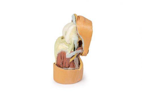 3D ANATOMY - Deep dissection of the knee joint, flexed
See details →
-
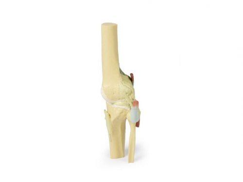 3D ANATOMY - Knee joint, extended
See details →
-
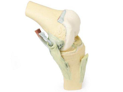 3D ANATOMY - Knee joint, flexed
See details →
-
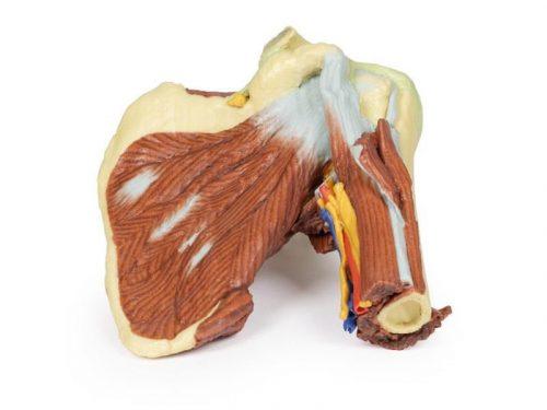 3D ANATOMY - Left shoulder, deep dissection of the joint, musculature, nerves and vessels
See details →
-
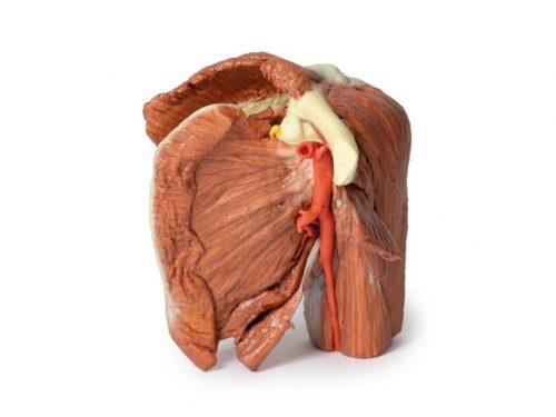 3D ANATOMY - Left shoulder, superficial muscles and axillary/brachial artery
See details →
-
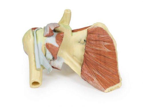 3D ANATOMY - Right shoulder, deep dissection of shoulder girdle, lateral clavicle and humerus
See details →
Is this equipment going into a new centre?
If you're equipping a classroom or a whole centre, starting from the shopping list works out expensive. Start from the needs assessment.