

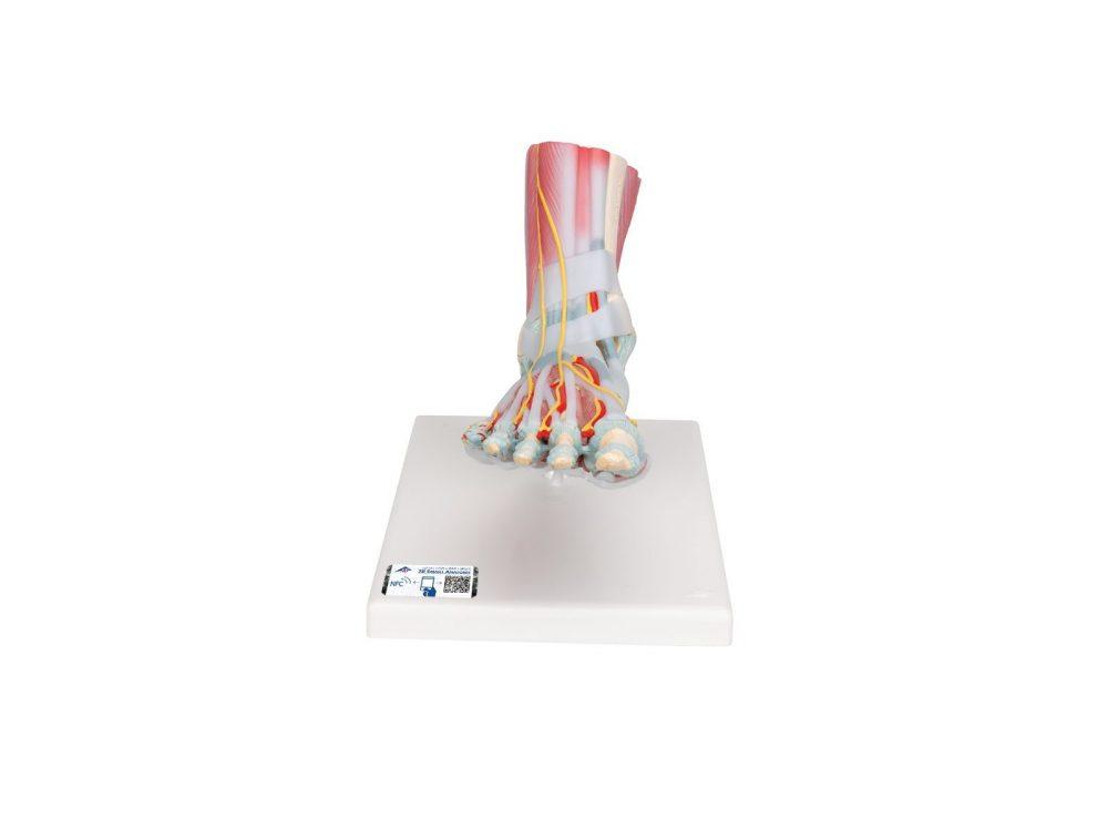
Reference 02191 · Anatomical model
Foot skeleton model with ligaments and muscles
- Anatomical model
- Orthopaedic surgery
- Main product
- Medicine
This anatomically detailed model of the foot and lower leg can be disassembled into 6 removable parts for detailed study of the foot and ankle. The foot skeleton features not only the bones but also the muscles, tendons, ligaments, nerves, arteries of the foot.
Price on request
Available in Spain, Portugal and Andorra · delivery time given in the proposal
Downloadable data sheet — features, specifications and delivery time, in PDF.
Need a technical report? Ask us for it and we'll send it within 24 h →
Description
What this equipment is and what it's designed for.
This anatomically detailed model of the foot and lower leg can be disassembled into 6 removable parts for detailed study of the foot and ankle. The foot skeleton features not only the bones but also the muscles, tendons, ligaments, nerves, arteries of the foot. The frontal view of the foot model features the extensor muscles of the lower leg. The tendons can be followed on their passage under the transverse and crucial crural ligaments all the way to their insertion points. In addition all tendon sheaths of the foot area are visible. On the dorsal portion of the foot the gastrocnemius muscle is removable to reveal deeper anatomical elements. The sole of the foot is represented in three layers; the first layer displaying the flexor digitorum brevis. This muscle can be removed from the foot revealing the quadratus plantae, the tendon of the flexor digitorum longus, and the flexor hallucis muscle. This second layer is in turn removable to display even deeper anatomical details of the foot. This foot skeleton model with ligaments and muscles is the best of its kind in quality and value.
What you learn
Anatomical model modality. The competencies this equipment trains, in the language of the training plan.
- Identification of bones, muscles, tendons, ligaments, nerves and arteries of the foot and the leg
- Recognition of the extensor muscles of the leg and of their tendon sheaths
- Layer-by-layer study of the sole of the foot
- Location of the tendon insertion points
- Description of the deep plantar musculature
Features and what's included
What the equipment does and what comes in the box, as described by the manufacturer.
Features
- Model of the foot and the leg, dismantles into 6 parts
- It shows bones, muscles, tendons, ligaments, nerves and arteries
- Front view with the extensor muscles and the tendon sheaths visible
- Removable calf muscle on the posterior aspect
- Sole of the foot represented in three layers, the first two removable
Services for this equipment
Services matter: the equipment gets commissioned, the staff know how to use it and maintenance is settled before you start. Include them in your budget and avoid having to raise a second purchase.
- Preventive maintenance We'll let you know when it's due
Documentation
You can download it now or we'll send it on request.
Other related equipment
They share a specialty with this one. They train different things: none of them replaces it.
-
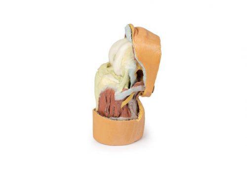 3D ANATOMY - Deep dissection of the knee joint, flexed
See details →
-
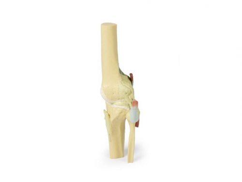 3D ANATOMY - Knee joint, extended
See details →
-
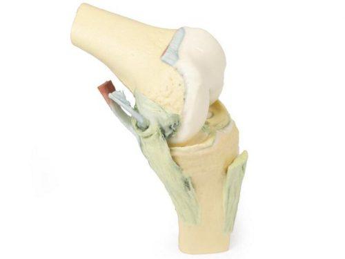 3D ANATOMY - Knee joint, flexed
See details →
-
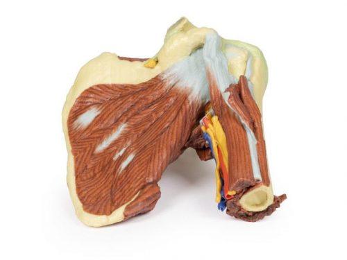 3D ANATOMY - Left shoulder, deep dissection of the joint, musculature, nerves and vessels
See details →
-
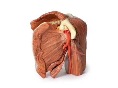 3D ANATOMY - Left shoulder, superficial muscles and axillary/brachial artery
See details →
-
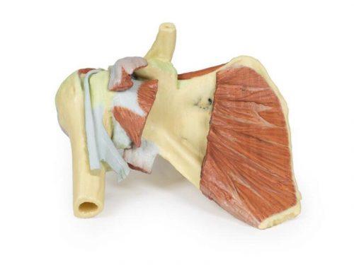 3D ANATOMY - Right shoulder, deep dissection of shoulder girdle, lateral clavicle and humerus
See details →
Is this equipment going into a new centre?
If you're equipping a classroom or a whole centre, starting from the shopping list works out expensive. Start from the needs assessment.