

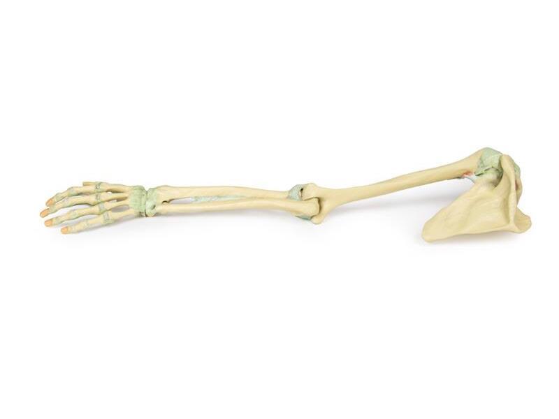
Reference 02686 · Anatomical model
3D ANATOMY - Upper limb ligaments
- Anatomical model
- Orthopaedic surgery
- Main product
- Medicine
Complete upper limb skeleton, from the pectoral girdle to the hand, with detailed ligamentous anatomy.
Price on request
Available in Spain, Portugal and Andorra · delivery time given in the proposal
Downloadable data sheet — features, specifications and delivery time, in PDF.
Need a technical report? Ask us for it and we'll send it within 24 h →
Description
What this equipment is and what it's designed for.
This 3D printed model presents the entire upper limb skeleton, from the pectoral girdle to the hand, with detailed ligamentous anatomy and select tendon and muscle insertions.
Shoulder and Pectoral Girdle
Visible ligaments include the acromioclavicular, coracoclavicular, coracoacromial, and superior transverse scapular ligament. A small portion of the supraspinatus muscle and tendon demonstrates its path beneath the coracoacromial ligament.
Key preserved structures:
– Subscapularis tendon (reflected to expose the glenohumeral capsule)
– Tendons of teres major, latissimus dorsi, and long head of triceps
– Long head of biceps tendon within the intertubercular groove and capsule
Elbow Joint
The elbow capsule is opened to show the articulation between the humerus, radius, and ulna.
Preserved:
– Ulnar and radial collateral ligaments
– Anular ligament of radius
– Biceps tendon insertion on the radial tuberosity
Wrist and Hand
Distally, the model includes key wrist ligaments:
– Palmar/dorsal radiocarpal and ulnocarpal ligaments
– Radial/ulnar collateral ligaments, pisohamate, carpometacarpal, and others
In the hand:
– MCP and IP joint capsules with collateral ligaments and volar plates
– Flexor digitorum superficialis/profundus and flexor pollicis longus tendons are preserved and shown at their insertions
What you learn
Anatomical model modality. The competencies this equipment trains, in the language of the training plan.
- Identify the ligaments of the shoulder, elbow, wrist and hand.
- Relate each ligament to the movement it restrains.
- Explain the injury mechanism of dislocations and sprains.
- Support joint stability testing on the model.
Cases and pathologies
The clinical scenarios that can be simulated with this equipment.
- Glenohumeral dislocation and capsuloligamentous injury.
- Sprain of the ulnar collateral ligament of the thumb.
- Carpal instability.
- Elbow dislocation.
Features and what's included
What the equipment does and what comes in the box, as described by the manufacturer.
Features
- 3D printed replica made from radiological data or from a dissected cadaveric specimen, not a stylised model: this is real anatomy.
- Developed and checked by the anatomists of the Human Anatomy Teaching Centre at Monash University.
- Colourised to distinguish muscle, nerve, artery, vein and bone, and to separate territories or segments.
- Not human tissue: no restrictions on transport, import or use in institutions without permission to work with cadavers.
- Avoids the ethical and preservation problems that come with plastination.
- Supplied with a detailed description of the anatomy represented in the specimen.
Services for this equipment
Services matter: the equipment gets commissioned, the staff know how to use it and maintenance is settled before you start. Include them in your budget and avoid having to raise a second purchase.
- Preventive maintenance We'll let you know when it's due
Documentation
You can download it now or we'll send it on request.
Other related equipment
They share a specialty with this one. They train different things: none of them replaces it.
-
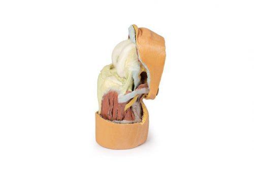 3D ANATOMY - Deep dissection of the knee joint, flexed
See details →
-
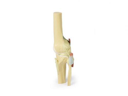 3D ANATOMY - Knee joint, extended
See details →
-
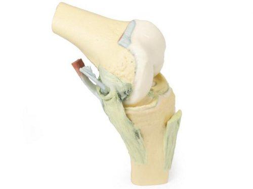 3D ANATOMY - Knee joint, flexed
See details →
-
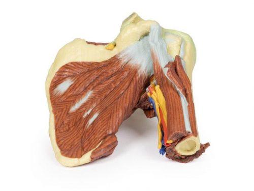 3D ANATOMY - Left shoulder, deep dissection of the joint, musculature, nerves and vessels
See details →
-
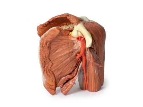 3D ANATOMY - Left shoulder, superficial muscles and axillary/brachial artery
See details →
-
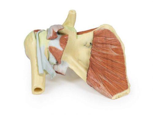 3D ANATOMY - Right shoulder, deep dissection of shoulder girdle, lateral clavicle and humerus
See details →
Is this equipment going into a new centre?
If you're equipping a classroom or a whole centre, starting from the shopping list works out expensive. Start from the needs assessment.