

Reference 02633 · Anatomical model
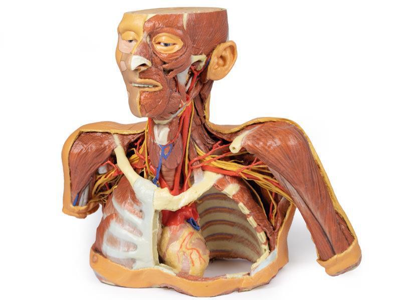
3D ANATOMY - Head, neck and shoulder with angiosomes
- Anatomical model
- Anatomy
- Main product
- Medicine
Multipart specimen of head, neck, thorax, axillae and upper limbs, with the angiosomes colour-coded.
Price on request
Available in Spain, Portugal and Andorra · delivery time given in the proposal
Downloadable data sheet — features, specifications and delivery time, in PDF.
Need a technical report? Ask us for it and we'll send it within 24 h →
Description
What this equipment is and what it's designed for.
This large, multipart 3D printed specimen showcases detailed anatomy of the head, neck, thorax, axillae, and upper limbs.
Head and Neck:
The calotte has been removed ~2?cm above the orbits to expose the brain and endocranial cavity. A transverse cerebral section shows grey and white matter, lateral ventricles, and choroid plexus. The right side retains skin and fascia, false-coloured to highlight facial and neck angiosomes. The left side reveals facial expression and mastication muscles, and infratemporal structures including the lingual nerve and terminal branches of the external carotid artery. The carotid sheaths are opened bilaterally, exposing the common, internal, and external carotid arteries, and vagus nerves. The sternocleidomastoid and internal jugular veins are mostly removed. On the right, the great auricular and hypoglossal nerves are visible, along with the stylohyoid ligament and supra-/infrahyoid muscles. The thyroid gland is prominent, with preserved superior and inferior thyroid vessels.
Root of the Neck and Axilla:
On the left, partial clavicle removal reveals the first rib, anterior scalene, and brachial plexus roots (C5–T1) forming trunks between scalene muscles. The subclavian artery passes posterior to scalenus anterior, transitioning to the axillary artery, closely related to the brachial plexus cords.
The left axilla displays brachial plexus divisions and cords. The formation of the median nerve around the axillary artery is distinct. The ulnar, musculocutaneous, axillary, thoracodorsal, and long thoracic nerves are clearly identified with their courses and muscular targets.
On the right, the clavicle and subclavius muscle are intact, showing the cervico-axillary canal. Pectoralis major and minor have been reflected, exposing deeper structures.
Thorax:
A window in the left thoracic wall reveals the mediastinum. The left lung has been removed. Intercostal spaces are visible beneath the parietal pleura; neurovascular bundles are identifiable posteriorly. The heart is exposed without pericardium, showing the left atrium and ventricle, pulmonary vessels, aorta, and both left vagus and recurrent laryngeal nerves. The right thoracic wall remains intact, displaying intercostal and upper limb muscles. From below, the right lung, pleural cavities, and diaphragmatic heart surface are visible. Posterior thoracic skin and fascia are intact, showing cutaneous nerve distribution.
Size: 50 x 20 x 41 cm
What you learn
Anatomical model modality. The competencies this equipment trains, in the language of the training plan.
- Identify the vascular territories (angiosomes) of the head, neck and shoulder.
- Follow the carotid arteries and their branches to each territory.
- Relate the arterial supply to flap design in reconstructive surgery.
- Place the surface landmarks of the neck over the deep structures.
Cases and pathologies
The clinical scenarios that can be simulated with this equipment.
- Flap planning in head and neck reconstructive surgery.
- Ischaemic compromise of a cutaneous territory.
- Cervical approaches with vascular risk.
- Teaching vascular anatomy for surgery and radiology.
Features and what's included
What the equipment does and what comes in the box, as described by the manufacturer.
Features
- 3D printed replica made from radiological data or from a dissected cadaveric specimen, not a stylised model: this is real anatomy.
- Developed and checked by the anatomists of the Human Anatomy Teaching Centre at Monash University.
- Colourised to distinguish muscle, nerve, artery, vein and bone, and to separate territories or segments.
- Not human tissue: no restrictions on transport, import or use in institutions without permission to work with cadavers.
- Avoids the ethical and preservation problems that come with plastination.
- Supplied with a detailed description of the anatomy represented in the specimen.
Services for this equipment
Services matter: the equipment gets commissioned, the staff know how to use it and maintenance is settled before you start. Include them in your budget and avoid having to raise a second purchase.
- Preventive maintenance We'll let you know when it's due
Documentation
You can download it now or we'll send it on request.
Other related equipment
They share a specialty with this one. They train different things: none of them replaces it.
-
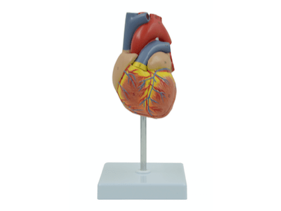 2-Part Deluxe Life-Size Human Heart Anatomy Model
See details →
-
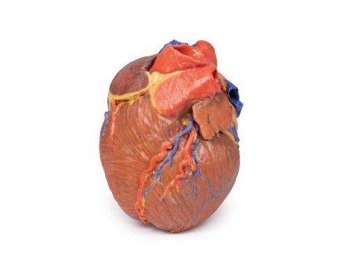 3D ANATOMY - Complete heart in 3D
See details →
-
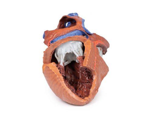 3D ANATOMY - Internal structures of the heart
See details →
-
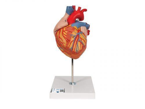 Anatomical heart model, x2 Magnification - 4 Parts
See details →
-
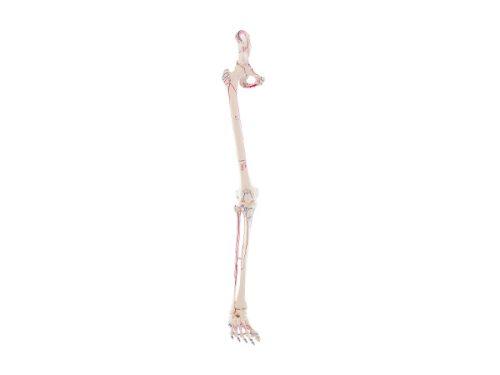 Anatomical model of artery and vein structure, x20 magnification
See details →
-
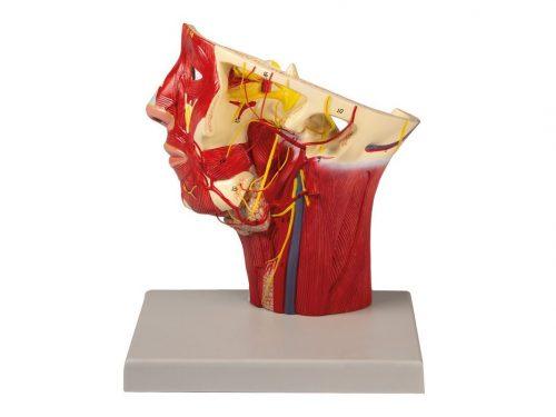 Arteries of head
See details →
Is this equipment going into a new centre?
If you're equipping a classroom or a whole centre, starting from the shopping list works out expensive. Start from the needs assessment.