

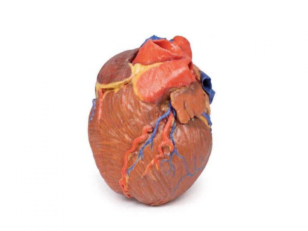
Reference 02693 · Anatomical model
3D ANATOMY - Complete heart in 3D
- Anatomical model
- Anatomy
- Main product
- Medicine
Isolated human heart with the superficial anatomy and the bases of the great vessels.
Price on request
Available in Spain, Portugal and Andorra · delivery time given in the proposal
Downloadable data sheet — features, specifications and delivery time, in PDF.
Need a technical report? Ask us for it and we'll send it within 24 h →
Description
What this equipment is and what it's designed for.
This 3D print preserves the superficial anatomy of an isolated human heart along with the bases of the great vessels. All four chambers—atria and ventricles—are clearly visible, with pericardial reflections on the left atrium marking the positions of the transverse and oblique pericardial sinuses.
Coronary Arteries and Veins
The right marginal branch of the right coronary artery is visible exiting the fat-filled coronary sulcus, along with the posterior interventricular (posterior descending) artery within its sulcus. On the anterior side, the anterior interventricular (left anterior descending) artery and diagonal branches from the left coronary artery are clearly displayed. The terminal portion of the circumflex branch can be seen deep to the left auricle, alongside the great cardiac vein. On the posterior aspect, the coronary sinus collects all cardiac veins—great, middle, and small—and includes a prominent posterior vein of the left ventricle.
Heart Valves
At the bases of the ascending aorta and pulmonary trunk, the aortic and pulmonary semilunar valves are visible, providing a detailed view of the outflow pathways from the ventricles.
What you learn
Anatomical model modality. The competencies this equipment trains, in the language of the training plan.
- Identify the cardiac chambers, the valves and the great vessels.
- Follow the course of the coronary arteries over the surface of the heart.
- Relate the anatomy to the auscultation areas.
- Orient the heart as it appears on chest radiography and echocardiography.
Cases and pathologies
The clinical scenarios that can be simulated with this equipment.
- Ischaemic heart disease by affected coronary artery.
- Valve disease and its murmurs.
- Heart failure and chamber remodelling.
- Teaching cardiac auscultation and examination.
Features and what's included
What the equipment does and what comes in the box, as described by the manufacturer.
Features
- 3D printed replica made from radiological data or from a dissected cadaveric specimen, not a stylised model: this is real anatomy.
- Developed and checked by the anatomists of the Human Anatomy Teaching Centre at Monash University.
- Colourised to distinguish muscle, nerve, artery, vein and bone, and to separate territories or segments.
- Not human tissue: no restrictions on transport, import or use in institutions without permission to work with cadavers.
- Avoids the ethical and preservation problems that come with plastination.
- Supplied with a detailed description of the anatomy represented in the specimen.
Services for this equipment
Services matter: the equipment gets commissioned, the staff know how to use it and maintenance is settled before you start. Include them in your budget and avoid having to raise a second purchase.
- Preventive maintenance We'll let you know when it's due
Documentation
You can download it now or we'll send it on request.
Other related equipment
They share a specialty with this one. They train different things: none of them replaces it.
-
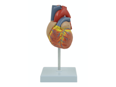 2-Part Deluxe Life-Size Human Heart Anatomy Model
See details →
-
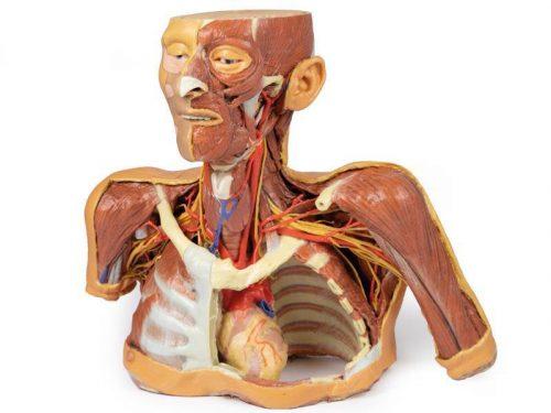 3D ANATOMY - Head, neck and shoulder with angiosomes
See details →
-
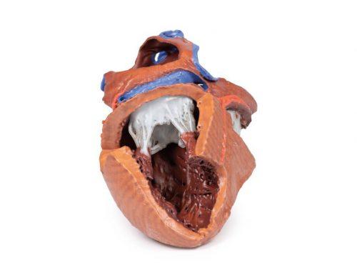 3D ANATOMY - Internal structures of the heart
See details →
-
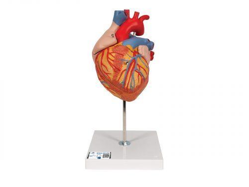 Anatomical heart model, x2 Magnification - 4 Parts
See details →
-
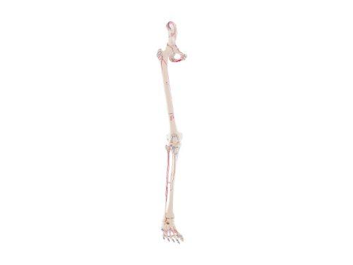 Anatomical model of artery and vein structure, x20 magnification
See details →
-
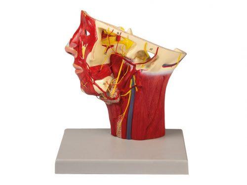 Arteries of head
See details →
Is this equipment going into a new centre?
If you're equipping a classroom or a whole centre, starting from the shopping list works out expensive. Start from the needs assessment.