

Reference 02702 · Anatomical model
3D ANATOMY - Superficial and deep structures in female pelvis
- Anatomical model
- Skeletal system
- Main product
- Medicine
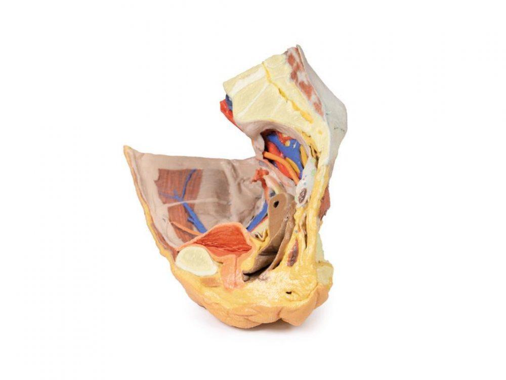
True and false female pelvis with inguinal ligament, obturator canal and sciatic foramina, and windows onto the extraperitoneal structures.
Price on request
Available in Spain, Portugal and Andorra · delivery time given in the proposal
Downloadable data sheet — features, specifications and delivery time, in PDF.
Need a technical report? Ask us for it and we'll send it within 24 h →
Description
What this equipment is and what it's designed for.
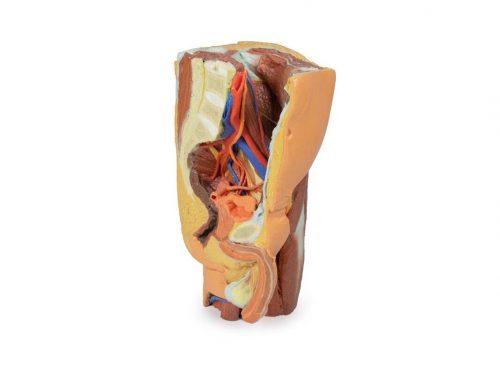
This 3D printed specimen preserves superficial and deep structures of the true and false pelvis, the inguinal ligament, obturator canal, and sciatic foramina, with ‘windows’ exposing extraperitoneal structures.
Posterior Abdominal Wall & Major Vessels
Sectioned at L4, it shows the colon, psoas and quadratus lumborum, and abdominal wall muscles. The common iliac artery and vein, inferior vena cava, and the bifurcation into external and internal iliac arteries are clearly visible. The external iliac vessels pass along the pelvic brim, giving off inferior epigastric and deep circumflex branches, with the femoral nerve lateral to psoas and the lateral cutaneous nerve of the thigh running over iliacus.
Internal Iliac & Obturator Structures
Branches of the internal iliac artery supply the bladder, uterus, and lateral pelvic wall, while the obturator nerve, artery, and vein pass through the obturator canal. Sacral nerves (S1–S3) form the sciatic nerve, which exits via the greater sciatic foramen alongside gluteal vessels.
Pelvic Organs
The ureter, bladder, uterus, vagina, and cervix are clearly visible in midsagittal view. The Fallopian tube, fimbria, and ovary with its ligaments are preserved, as well as small portions of the rectum and nearby pararectal lymph nodes.
Femoral Triangle & Gluteal Region
The inguinal ligament is intact, with femoral vessels and nerve passing beneath it. In the gluteal region, the sciatic nerve and gluteal vessels are visible, and the pudendal nerve and vessels pass into the lesser sciatic foramen toward the perineum.
What you learn
Anatomical model modality. The competencies this equipment trains, in the language of the training plan.
- Compare the superficial and the deep plane of the female pelvis.
- Identify the pelvic floor and the organs it supports.
- Recognise the relation between bladder, uterus and rectum.
- Relate the anatomy to gynaecological examination and to childbirth.
Cases and pathologies
The clinical scenarios that can be simulated with this equipment.
- Pelvic organ prolapse.
- Stress urinary incontinence.
- Perineal tear during delivery.
- Gynaecological surgery and its dissection planes.
Features and what's included
What the equipment does and what comes in the box, as described by the manufacturer.
Features
- 3D printed replica made from radiological data or from a dissected cadaveric specimen, not a stylised model: this is real anatomy.
- Developed and checked by the anatomists of the Human Anatomy Teaching Centre at Monash University.
- Colourised to distinguish muscle, nerve, artery, vein and bone, and to separate territories or segments.
- Not human tissue: no restrictions on transport, import or use in institutions without permission to work with cadavers.
- Avoids the ethical and preservation problems that come with plastination.
- Supplied with a detailed description of the anatomy represented in the specimen.
Services for this equipment
Services matter: the equipment gets commissioned, the staff know how to use it and maintenance is settled before you start. Include them in your budget and avoid having to raise a second purchase.
- Preventive maintenance We'll let you know when it's due
Documentation
You can download it now or we'll send it on request.
Other related equipment
They share a specialty with this one. They train different things: none of them replaces it.
Is this equipment going into a new centre?
If you're equipping a classroom or a whole centre, starting from the shopping list works out expensive. Start from the needs assessment.