

Reference 10994 · Anatomical model
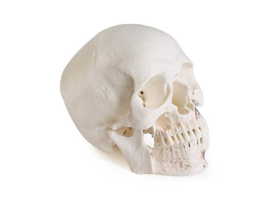
Skull model for dentistry with TMD syndrome, 8 parts
- Anatomical model
- Anatomy
- Main product
- Dentistry
In addition to the detailed reproduction of the skull structures, this first-class model of a human skull focuses specifically on teeth and jaws and is therefore the perfect teaching and demonstration tool for the field of dentistry and oral surgery. The front half of the upper and lower jaw can be removed to reveal t
Price on request
Available in Spain, Portugal and Andorra · delivery time given in the proposal
Downloadable data sheet — features, specifications and delivery time, in PDF.
Need a technical report? Ask us for it and we'll send it within 24 h →
Description
What this equipment is and what it's designed for.
In addition to reproducing the structures of the skull in detail, this premium-quality human skull model focuses especially on the teeth and the jaws, which makes it ideal for teaching and demonstration in dentistry and oral surgery.
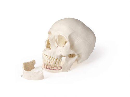
The anterior half of the maxilla and of the mandible can be removed to expose the teeth with their roots, the nerves and the blood vessels on one side and the maxillary sinuses. The skull serves both as support for learning anatomy and as an aid for explaining oral surgery and implantology.
This special design serves to illustrate temporomandibular dysfunction. In temporomandibular dysfunction there is a disturbance in the interaction between the temporomandibular joints, the masticatory muscles and the teeth. The wear of the dental enamel from bruxism, the wear of the disc and the masticatory muscles involved are shown: the masseter, in its pars superficialis and its pars profundus, and the pars coronoidea, which was discovered relatively recently. All three muscles are removable.
What you learn
Anatomical model modality. The competencies this equipment trains, in the language of the training plan.
- Identificar los dientes, sus raíces y el proceso alveolar.
- Reconocer los músculos masticatorios implicados en la disfunción temporomandibular.
- Explicar la interacción entre articulación temporomandibular, músculos y dientes.
- Mostrar los signos del bruxismo sobre el esmalte y el disco articular.
Cases and pathologies
The clinical scenarios that can be simulated with this equipment.
- Disfunción temporomandibular.
- Bruxismo y desgaste del esmalte dental.
- Desgaste del disco articular.
- Docencia de odontología, cirugía oral e implantología.
Features and what's included
What the equipment does and what comes in the box, as described by the manufacturer.
Features
- Modelo de cráneo humano de 8 partes, centrado en dientes y maxilares.
- Mitad anterior del maxilar y de la mandíbula extraíble, con raíces, nervios, vasos y senos maxilares a la vista.
- Representa el masetero (pars superficialis y pars profundus) y la pars coronoidea.
- Los tres músculos masticatorios son desmontables.
Services for this equipment
Services matter: the equipment gets commissioned, the staff know how to use it and maintenance is settled before you start. Include them in your budget and avoid having to raise a second purchase.
- Preventive maintenance We'll let you know when it's due
Documentation
You can download it now or we'll send it on request.
Other related equipment
They share a specialty with this one. They train different things: none of them replaces it.
Is this equipment going into a new centre?
If you're equipping a classroom or a whole centre, starting from the shopping list works out expensive. Start from the needs assessment.