

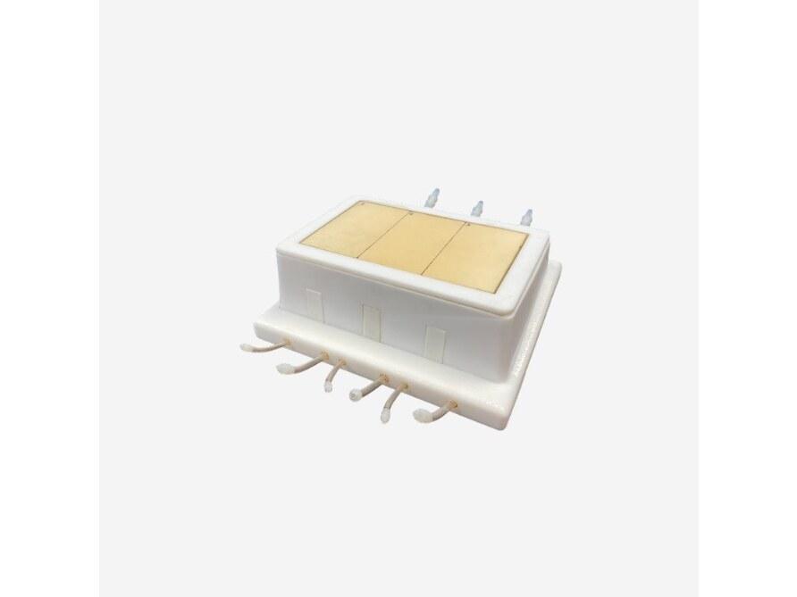
Reference 10405 · Task trainer
Doppler Phantom for X-Ray CT, US, MRI
- Task trainer
- Angiology
- Main product
- Medicine
Doppler Phantom serves purposes in medical imaging research, development, training, and testing. The phantom includes a bone plate simulating the 2D projection of the skull with realistic internal porosity.
Price on request
Available in Spain, Portugal and Andorra · delivery time given in the proposal
Downloadable data sheet — features, specifications and delivery time, in PDF.
Need a technical report? Ask us for it and we'll send it within 24 h →
Description
What this equipment is and what it's designed for.
Doppler Phantoms are used for medical imaging research and development, training, and testing purposes. The bone plate installed on the top of this phantom mimics the skull bone, which in this case simulates only the 2D projection of the skull with a realistic internal porosity structure. The internal blood vessels can be filled either with water or with most of the commonly used blood-mimicking fluids. This phantom can be used with Barium-sulfate, Iodine-based, Gadolinium – based, Microbubble, and Iron Oxide-based contrast mediums.
The phantom can be customized in multiple ways to suit the needs of a wide variety of applications.
Size: 8 x 6 x 4 Inches, Weight: 1 Lbs (approx.).
Size: 12 x 10 x 10 Inches, Weight: 6 Lbs (approx.)
What you learn
Task trainer modality. The competencies this equipment trains, in the language of the training plan.
- Adjust the insonation angle, the PRF and the gain until a valid waveform is obtained.
- Position and centre the specimen for each projection or slice plane.
- Choose the radiographic technique and check its effect on the image.
- Adjust the CT protocol and assess the result on the reconstructions.
- Orient the probe and recognise the structures in each ultrasound plane.
- Try out MRI sequences and compare the contrast between tissues.
Related procedures: MRI · Tomography · Ultrasound · X-ray
Cases and pathologies
The clinical scenarios that can be simulated with this equipment.
- Setting up colour Doppler and pulsed Doppler.
- Quality control of velocity measurement.
- Training in the study of arterial and venous flow.
- Acceptance testing of a unit or validation of a new protocol.
- Training radiographers and residents without exposing any patient.
Features and what's included
What the equipment does and what comes in the box, as described by the manufacturer.
Features
- Imaging response equivalent to human tissue in X-ray, CT, ultrasound and MRI.
- Differentiated internal anatomy, not a homogeneous block.
- No ferromagnetic components: it can enter the MRI room.
- Takes direct probe contact with gel.
- It can be scanned as often as needed and exposes nobody to radiation.
Includes
- User Manual/Assembly Instructions
- 2-Year Warranty & Unlimited Customer Service
Services for this equipment
Services matter: the equipment gets commissioned, the staff know how to use it and maintenance is settled before you start. Include them in your budget and avoid having to raise a second purchase.
- Preventive maintenance We'll let you know when it's due
Documentation
You can download it now or we'll send it on request.
Other related equipment
They train the same technique. Each one approaches it in its own way: none of them replaces this one.
-
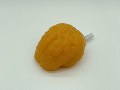 Adult Brain (Standard) for X-Ray CT, US, MRI
See details →
-
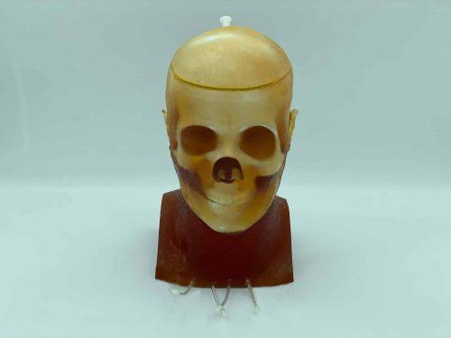 Adult Head (Dynamic, Advanced) for X-Ray CT, US, MRI
See details →
-
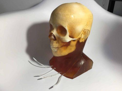 Adult Head (Dynamic) for X-Ray CT, US, MRI
See details →
-
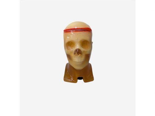 Adult Head (Openable) for X-Ray CT, US, MRI
See details →
-
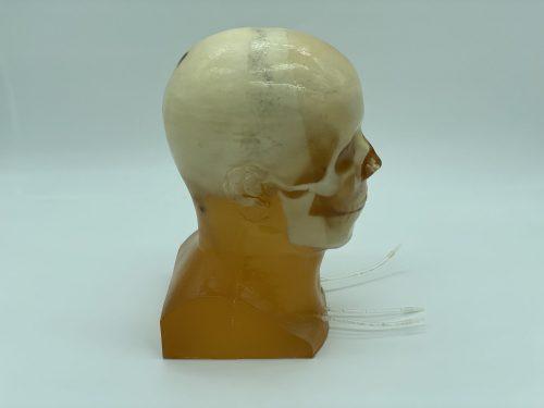 Adult Head (Standard) for X-Ray CT, US, MRI
See details →
-
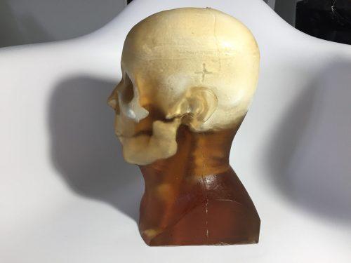 Adult Head (Static) for X-Ray CT, US, MRI
See details →
Is this equipment going into a new centre?
If you're equipping a classroom or a whole centre, starting from the shopping list works out expensive. Start from the needs assessment.