

Reference 14445 · Anatomical model
Canine Thoracic Cavity
- Anatomical model
- Anatomy
- Main product
- Veterinary medicine
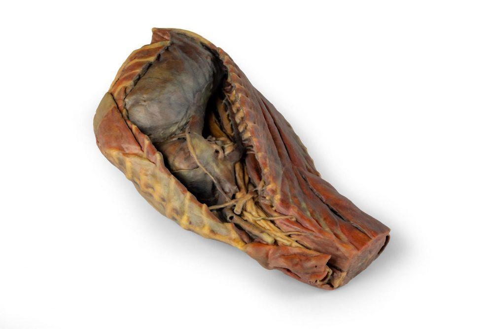
Right-side dissection of the thoracic cavity of an adult dog, with the thoracic wall and right lung removed.
Price on request
Available in Spain, Portugal and Andorra · delivery time given in the proposal
Downloadable data sheet — features, specifications and delivery time, in PDF.
Need a technical report? Ask us for it and we'll send it within 24 h →
Description
What this equipment is and what it's designed for.
This specimen presents a right-side dissection of the thoracic cavity (Cavitas thoracis) of an adult dog. Following removal of the right thoracic wall, including the ribs (costae), the right lung (pulmo dexter) has been excised, leaving the pulmonary vessels (vasa pulmonalia) in situ in relation to the heart (cor). In the cranial mediastinum (mediastinum craniale), the veins forming the cranial vena cava (vena cava cranialis) are identifiable, along with the course of the branches of the right subclavian artery (arteria subclavia dextra). Also visible in this region are the thoracic portion of the longus colli muscle (musculus longus colli), the thoracic trachea (trachea thoracica) and the thoracic oesophagus (esophagus thoracicus).
The course of the phrenic nerve (nervus phrenicus) and the vagosympathetic trunk (truncus vagosympathicus) is evident, as is the location of the stellate ganglion (ganglion stellatum). In the middle mediastinum (mediastinum medium), the heart (cor), partially covered by the fibrous pericardium (pericardium fibrosum), is visible. At its base (basis cordis), the pulmonary vessels and the principal bronchi (bronchi principales), formed by the division of the trachea, can be observed.
The caudal mediastinum (mediastinum caudale) shows the course of the caudal vena cava (vena cava caudalis) and the right phrenic nerve (nervus phrenicus dexter), adjacent to the caudal mediastinal recess (recessus mediastini caudalis), which is defined by the fold of the caudal vena cava (plica venae cavae). The dorsal aspect is occupied by the oesophagus (esophagus), along which run the dorsal and ventral vagal trunks (truncus vagalis dorsalis et ventralis), as well as the thoracic aorta (aorta thoracica), the right azygos vein (vena azygos dextra) and the sympathetic trunk (truncus sympathicus).
Caudally, the diaphragm (diaphragma) has been preserved, showing its attachments to the costal arches (arcus costales) and lumbar vertebrae (vertebrae lumbales). Additionally, the extensor musculature of the cervical and thoracic spine (musculi extensores columnae vertebralis cervicalis et thoracicae), the intercostal muscles (musculi intercostales) and the brachial plexus (plexus brachialis) are retained.
What you learn
Anatomical model modality. The competencies this equipment trains, in the language of the training plan.
- Identify the contents of the canine mediastinum
- Follow the course of the phrenic and vagus nerves in the thorax
- Relate the heart to the great vessels, trachea and oesophagus
Cases and pathologies
The clinical scenarios that can be simulated with this equipment.
- Pleural effusion and thoracocentesis
- Megaoesophagus
- Mediastinal masses
Features and what's included
What the equipment does and what comes in the box, as described by the manufacturer.
Features
- Right-side dissection with thoracic wall and right lung removed
- Cranial, middle and caudal mediastinum with their vessels and nerves
- Heart with fibrous pericardium, pulmonary vessels and principal bronchi
- Diaphragm, intercostal muscles and brachial plexus preserved
Services for this equipment
Services matter: the equipment gets commissioned, the staff know how to use it and maintenance is settled before you start. Include them in your budget and avoid having to raise a second purchase.
- Preventive maintenance We'll let you know when it's due
Documentation
You can download it now or we'll send it on request.
Other related equipment
They share a specialty with this one. They train different things: none of them replaces it.
Is this equipment going into a new centre?
If you're equipping a classroom or a whole centre, starting from the shopping list works out expensive. Start from the needs assessment.