

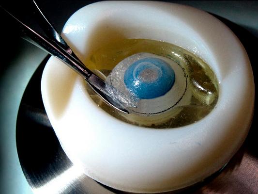
Reference 1674
BASIC ORBIT holder
- Ophthalmology
- Parts and Components
Basic Orbit for ALL anterior segment models in the range
Price on request
Available in Spain, Portugal and Andorra · delivery time given in the proposal
Downloadable data sheet — features, specifications and delivery time, in PDF.
Need a technical report? Ask us for it and we'll send it within 24 h →
Description
What this equipment is and what it's designed for.
The ORBIT is an anterior holder for BIONIKO anterior segment models. It provides the user with a compact yet realistic frame of reference, including anatomical landmarks such as the brow and eye movement simulation that provide an added challenge. This platform is a compromise between the MINI HOLDER and the FLEX-ORBIT.
Compatible with: OKULO, KERATO, RHEXIS, CORDELIA
What you learn
The competencies this equipment trains, in the language of the training plan.
Related procedures: Holders
Features and what's included
What the equipment does and what comes in the box, as described by the manufacturer.
Features
KEY FEATURES:- The ORBIT simulates the bony structure around the eye, including the nasal bones, the brow and cheekbone.The ORBIT has a flexible eyelid/socket (5) that receives and secures anterior segment models.TheORBITcan be secured to any smooth surface (horizontal or vertical) with its integrated suction cup and will still retain a realistic degree of freedom that simulates head and eye movement. There are both right (R) and left (L) ORBIT models to practice all approaches: Right-Superior, Right-Temporal, Left-Superior and Left-Temporal.
- Use a couple of water drops to moisten the eyelid and socket area. Insert the edge of the model under the superior eyelid (1) and push the other side down into the socket (2) to load the model. Be careful not to damage delicate structures when inserting the models. Always press on the sclera.
- Orient the ORBIT according to the desired approach. Fix the ORBIT in place by pressing downward on a smooth surface to engage the suction-cup.
- Remove model once a surgery has been practiced, remove the used model from the ORBIT by inserting a closed instrument in either corner of the eye and leveraging the model out (3). NOTE: Lift the suction release tab to remove ORBIT from surface. DO NOT PULL ON THE ORBIT
Includes
- 1 Basic orbit holder
Services for this equipment
Services matter: the equipment gets commissioned, the staff know how to use it and maintenance is settled before you start. Include them in your budget and avoid having to raise a second purchase.
- Preventive maintenance We'll let you know when it's due
Documentation
You can download it now or we'll send it on request.
Other related equipment
They share a specialty with this one. They train different things: none of them replaces it.
-
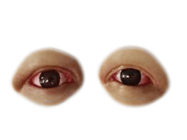 Conjunctival & Corneal Injuries
See details →
Conjunctival & Corneal Injuries
See details →
-
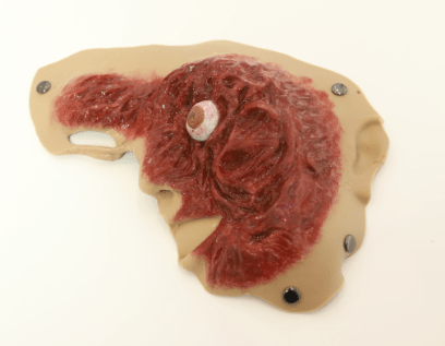 Eye Pop-out Injury
See details →
Eye Pop-out Injury
See details →
-
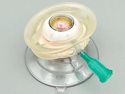 ANTERIOR VITRECTOMY KIT - Pre-assembled kit for anterior vitrectomy with empty capsule and rhexis (1 pc)
See details →
ANTERIOR VITRECTOMY KIT - Pre-assembled kit for anterior vitrectomy with empty capsule and rhexis (1 pc)
See details →
-
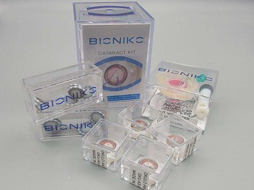 CATARACT KIT - Cataract surgery wet lab
See details →
CATARACT KIT - Cataract surgery wet lab
See details →
-
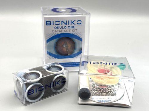 CATARACT OKULO ONE KIT - Cataract surgery starter kit with AVH holder and disposable anterior segment
See details →
CATARACT OKULO ONE KIT - Cataract surgery starter kit with AVH holder and disposable anterior segment
See details →
-
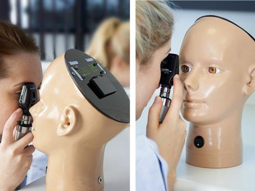 Digital eye examination trainer
See details →
Digital eye examination trainer
See details →
Is this equipment going into a new centre?
If you're equipping a classroom or a whole centre, starting from the shopping list works out expensive. Start from the needs assessment.