

Reference 03996 · Anatomical model
3D section model of the organ of Corti and cochlea, x350 magnification - 5 Parts
- Anatomical model
- Anatomy
- Main product
- Medicine
A three-dimensional model of the cochlear duct and the organ of Corti, magnified 350 times, for studying the histology of the inner ear.
Price on request
Available in Spain, Portugal and Andorra · delivery time given in the proposal
Downloadable data sheet — features, specifications and delivery time, in PDF.
Need a technical report? Ask us for it and we'll send it within 24 h →
Description
What this equipment is and what it's designed for.
Magnified 350 times life size and composed of 5 parts, this model shows a three-dimensional section of the cochlear duct, the tectorial membrane, the basilar membrane and the vestibular membrane.
The organ of Corti is represented in detail, including the acoustic receptor cells that convert sound vibrations into electrical signals.
Size: 47x18x32.5 cm. Weight: approx. 4500 g.
Services for this equipment
Services matter: the equipment gets commissioned, the staff know how to use it and maintenance is settled before you start. Include them in your budget and avoid having to raise a second purchase.
- Preventive maintenance We'll let you know when it's due
Documentation
You can download it now or we'll send it on request.
Other related equipment
They share a specialty with this one. They train different things: none of them replaces it.
-
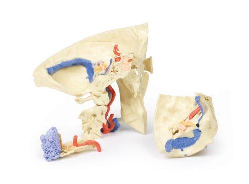 3D ANATOMY - Temporal bone model (Set of 3pcs)
See details →
-
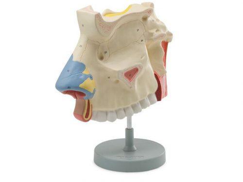 Anatomical model of the nasal cavity, x3 magnification - 2 Parts
See details →
-
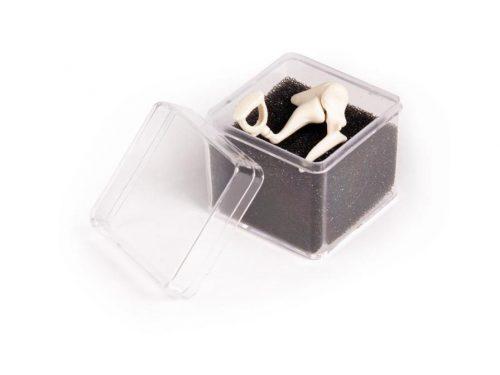 Auditory Ossicles, 3 times life size
See details →
-
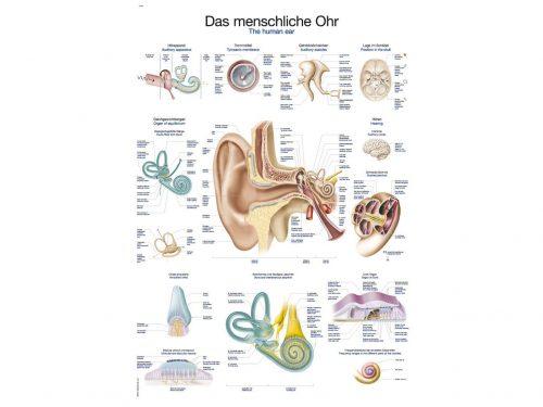 Chart The human ear, 50x70cm
See details →
-
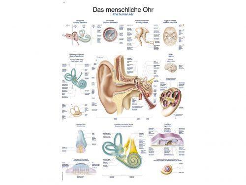 Chart The human ear, 70x100cm
See details →
-
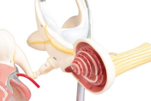 Ear labyrinth model with ossicles and eardrum
See details →
Is this equipment going into a new centre?
If you're equipping a classroom or a whole centre, starting from the shopping list works out expensive. Start from the needs assessment.