

Reference 02674 · Anatomical model
3D ANATOMY - Temporal bone model (Set of 3pcs)
- Anatomical model
- Anatomy
- Main product
- Medicine
Three-part temporal bone set derived from CT data: ossicles, canals, chambers, foramina and air cells.
Price on request
Available in Spain, Portugal and Andorra · delivery time given in the proposal
Downloadable data sheet — features, specifications and delivery time, in PDF.
Need a technical report? Ask us for it and we'll send it within 24 h →
Description
What this equipment is and what it's designed for.
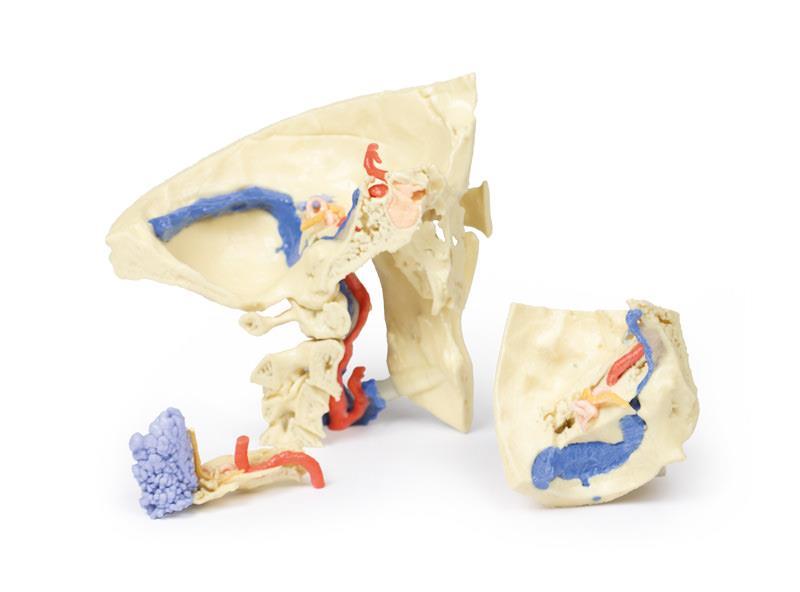
This 3-part 3D printed model, derived from CT data, highlights the complex anatomy of the temporal bone, including ossicles, canals, chambers, foramina, and air spaces. Internal casts (endocasts) reveal the intricate internal structure, aiding visualization of the auditory and vestibular apparatus.
Part 1: Skull Preparation
The model exposes the posterior quadrant of the skull, including the posterior and middle cranial fossa. The temporal bone and its relationship with the sphenoid, parietal, and occipital bones are clearly visible.
The middle ear (orange) shows the tympanum, aditus, antrum, and pharyngotympanic tube. The bony labyrinth of the inner ear (green) displays the semicircular canals and cochlea. The facial nerve (CN VII) is shown in yellow, tracing its course to the stylomastoid foramen. Mastoid air cells (blue) and the temporomandibular joint are also demonstrated.
The model highlights key vascular structures, including the internal carotid artery, transverse and sigmoidal dural venous sinuses, and the jugular foramen, along with the foramen magnum and first three cervical vertebrae.
Part 2: Petrous Part of the Temporal Bone
Enlarged x3, this section emphasizes the detailed internal architecture of the petrous temporal bone, including the middle ear ossicles (malleus, incus, stapes), mastoid air cells, and bony canal of the internal carotid artery.
The facial nerve and chorda tympani nerve are traced through the tympanic cavity, illustrating their relationship with the auditory structures. The model highlights connections between the tympanum and mastoid air cells via the aditus and antrum.
Part 3: Auditory and Vestibular Apparatus
Also enlarged x3, this model section focuses on the auditory and vestibular apparatus and their relation to surrounding otological structures. The bony labyrinth, ossicles, and tympanic connections to the nasopharynx are clearly shown.
The model demonstrates the passage of the facial (CN VII) and vestibulocochlear (CN VIII) nerves through the temporal bone, including the cochlear nerve, geniculate ganglion, and chorda tympani. The spatial relationship between these nerves, the internal carotid artery, and dural venous sinuses is accurately represented.
What you learn
Anatomical model modality. The competencies this equipment trains, in the language of the training plan.
- Orient the temporal bone and identify the middle ear, inner ear and mastoid air cells.
- Follow the intratemporal course of the facial nerve.
- Place the landmarks that guide a mastoidectomy.
- Repeat the exercise on several specimens until spatial orientation is secure.
Related procedures: Bone drilling and osteosynthesis
Cases and pathologies
The clinical scenarios that can be simulated with this equipment.
- Chronic otitis media and cholesteatoma.
- Mastoiditis.
- Cochlear implant surgery.
- Facial palsy of temporal origin.
Features and what's included
What the equipment does and what comes in the box, as described by the manufacturer.
Features
- 3D printed replica made from radiological data or from a dissected cadaveric specimen, not a stylised model: this is real anatomy.
- Developed and checked by the anatomists of the Human Anatomy Teaching Centre at Monash University.
- Colourised to distinguish muscle, nerve, artery, vein and bone, and to separate territories or segments.
- Not human tissue: no restrictions on transport, import or use in institutions without permission to work with cadavers.
- Avoids the ethical and preservation problems that come with plastination.
- Supplied with a detailed description of the anatomy represented in the specimen.
Services for this equipment
Services matter: the equipment gets commissioned, the staff know how to use it and maintenance is settled before you start. Include them in your budget and avoid having to raise a second purchase.
- Preventive maintenance We'll let you know when it's due
Documentation
You can download it now or we'll send it on request.
Other related equipment
They train the same technique. Each one approaches it in its own way: none of them replaces this one.
- BONE BLOCK - Solid bone block, 0.25g/cm3, 10x10x10cm See details →
- BONE BLOCK - Solid bone block, 0.25g/cm3, 15x15x15cm See details →
- BONE BLOCK - Solid bone block, 0.25g/cm3, 5x5x5cm See details →
- BONE BLOCK - Solid bone block, 0.35g/cm3, 10x10x10cm See details →
- BONE BLOCK - Solid bone block, 0.35g/cm3, 15x15x15cm See details →
- BONE BLOCK - Solid bone block, 0.35g/cm3, 5x5x5cm See details →
Is this equipment going into a new centre?
If you're equipping a classroom or a whole centre, starting from the shopping list works out expensive. Start from the needs assessment.