

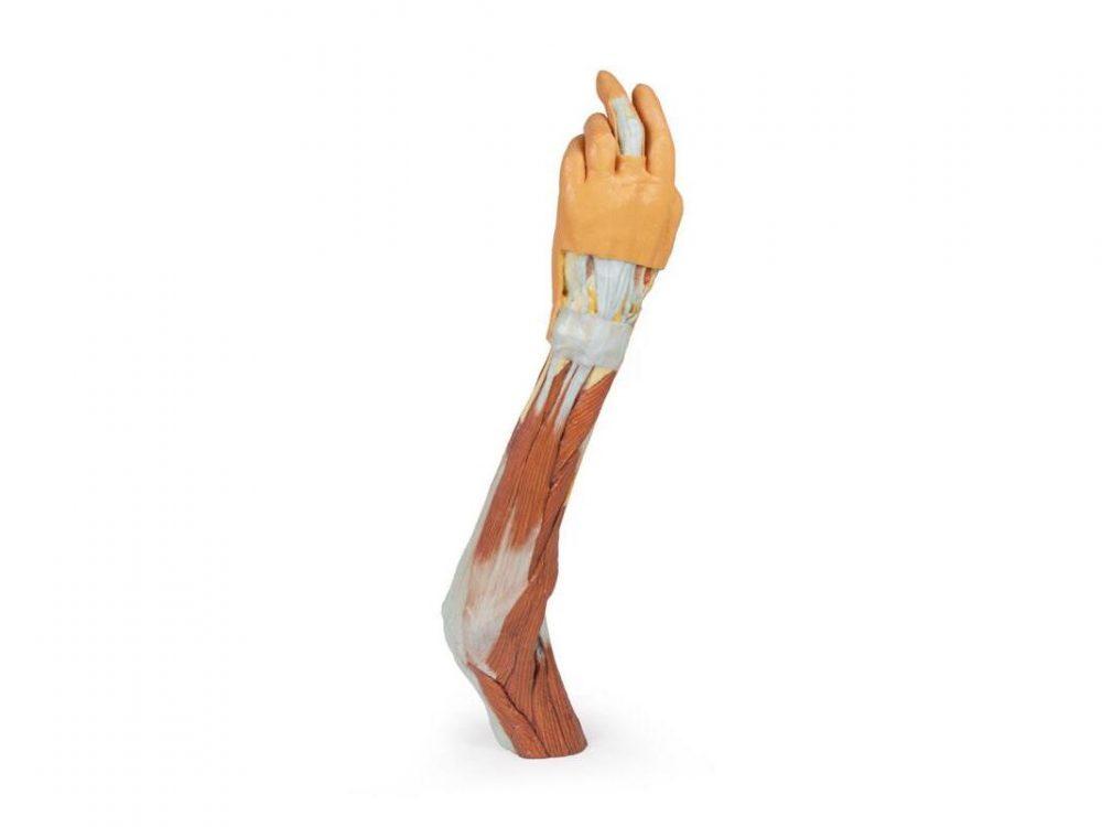
Reference 02681 · Anatomical model
3D ANATOMY - Upper limb (elbow, forearm and hand)
- Anatomical model
- Anatomy
- Main product
- Medicine
Left upper limb from the distal arm to the hand, in deep dissection of muscles, vessels and nerves.
Price on request
Available in Spain, Portugal and Andorra · delivery time given in the proposal
Downloadable data sheet — features, specifications and delivery time, in PDF.
Need a technical report? Ask us for it and we'll send it within 24 h →
Description
What this equipment is and what it's designed for.
This detailed 3D print displays the left upper limb from the distal arm to the hand, highlighting key muscular, vascular, and neural structures through deep dissection.
Elbow Region & Distal Arm
The cubital fossa reveals the classic lateral-to-medial layout: biceps tendon, brachial artery, and median nerve, with the bicipital aponeurosis removed. The ulnar nerve passes behind the medial epicondyle, and the superficial radial nerve is visible between brachioradialis and brachialis.
Anterior & Posterior Forearm
Anteriorly, the superficial flexors—pronator teres, FCR, FDS, and FCU—are clearly shown (no palmaris longus present). The radial and ulnar arteries, as well as the superficial radial nerve, are identifiable.
Posteriorly, extensor muscles from the common extensor origin are preserved, including ECU, extensor digitorum, and ECRB/ECRL. The anatomical snuffbox is visible with the radial artery and cutaneous radial nerve.
Palm & Carpal Tunnel
The thenar/hypothenar muscles, flexor retinaculum, long tendons, and lumbricals are exposed. The median nerve runs beneath the retinaculum, and the superficial palmar arch (from the ulnar artery) is intact. Digital nerves and vessels are clearly seen, along with the recurrent median nerve branch and the extensor expansion on the middle finger.
This model is ideal for studying upper limb anatomy, with special focus on the elbow, forearm, and hand structures.
What you learn
Anatomical model modality. The competencies this equipment trains, in the language of the training plan.
- Identify the elbow joint and the compartments of the forearm.
- Follow the median, ulnar and radial nerves to the hand.
- Locate the surface landmarks used for examination and for puncture.
- Relate the anatomy to fracture reduction and immobilisation.
Cases and pathologies
The clinical scenarios that can be simulated with this equipment.
- Elbow and forearm fractures.
- Lateral and medial epicondylitis.
- Ulnar neuropathy at the cubital tunnel.
- Forearm compartment syndrome.
Features and what's included
What the equipment does and what comes in the box, as described by the manufacturer.
Features
- 3D printed replica made from radiological data or from a dissected cadaveric specimen, not a stylised model: this is real anatomy.
- Developed and checked by the anatomists of the Human Anatomy Teaching Centre at Monash University.
- Colourised to distinguish muscle, nerve, artery, vein and bone, and to separate territories or segments.
- Not human tissue: no restrictions on transport, import or use in institutions without permission to work with cadavers.
- Avoids the ethical and preservation problems that come with plastination.
- Supplied with a detailed description of the anatomy represented in the specimen.
Services for this equipment
Services matter: the equipment gets commissioned, the staff know how to use it and maintenance is settled before you start. Include them in your budget and avoid having to raise a second purchase.
- Preventive maintenance We'll let you know when it's due
Documentation
You can download it now or we'll send it on request.
Other related equipment
They share a specialty with this one. They train different things: none of them replaces it.


Is this equipment going into a new centre?
If you're equipping a classroom or a whole centre, starting from the shopping list works out expensive. Start from the needs assessment.