

Reference 02177 · Anatomical model
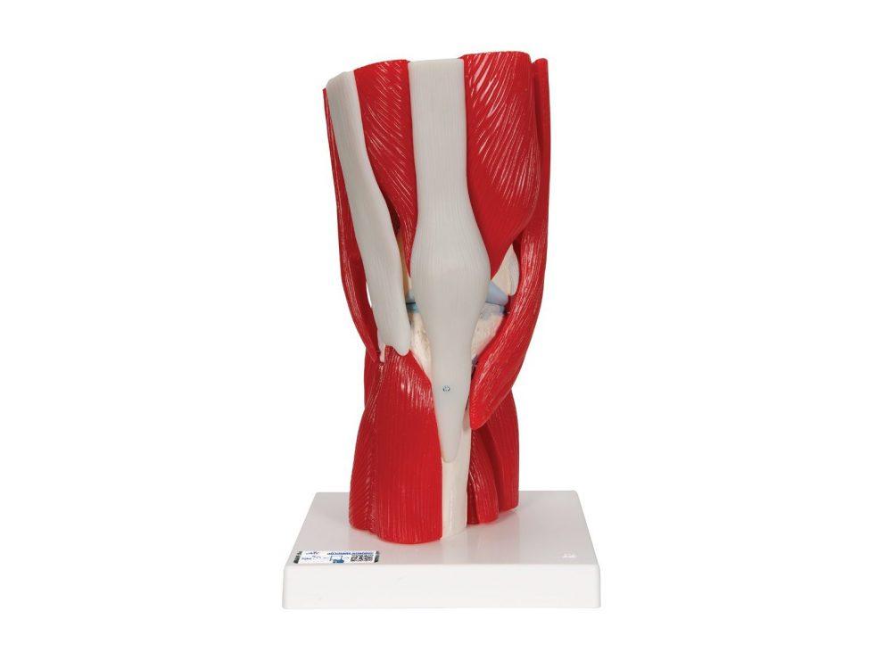
Knee joint model - 12 Parts
- Anatomical model
- Orthopaedic surgery
- Main product
- Medicine
The Knee Joint Model with removable joints is part of a high quality series of models. It has been manufactured to precise anatomical correctness with realistic colors of the joint, bone and muscles.
Price on request
Available in Spain, Portugal and Andorra · delivery time given in the proposal
Downloadable data sheet — features, specifications and delivery time, in PDF.
Need a technical report? Ask us for it and we'll send it within 24 h →
Description
What this equipment is and what it's designed for.
The Knee Joint Model with removable joints is part of a high quality series of models. It has been manufactured to precise anatomical correctness with realistic colors of the joint, bone and muscles. Only extremely durable, nonhazardous material of highest standards has been used throughout the production process.
The model consists of 12 parts which can be easily disassembled for easy demonstration. The muscles and muscle portions can be taken off, and a special color coding has been used to show muscle origin and insertion points on the femur, tibia, and fibula. Additionally, the model also shows parts of the fibular and tibial collateral ligaments.
Whether your are studying or teaching the deeper anatomical layers of the human knee joint, or are looking for a tool to better educate your patients about their injuries and rehab, the Knee Joint Model is an extraordinary teaching tool and offers exceptional value.
What you learn
Anatomical model modality. The competencies this equipment trains, in the language of the training plan.
- Identification of the muscles of the knee region
- Location of the points of origin and insertion on the femur, tibia and fibula
- Recognition of the tibial and fibular collateral ligaments
- Layer-by-layer study by removing the musculature
Features and what's included
What the equipment does and what comes in the box, as described by the manufacturer.
Features
- 12-part model of the knee joint
- Removable muscles and muscle portions
- Origin and insertion areas in relief and colour-coded
- It represents parts of the fibular and tibial collateral ligaments
- It allows the study of the deep anatomical layers
Services for this equipment
Services matter: the equipment gets commissioned, the staff know how to use it and maintenance is settled before you start. Include them in your budget and avoid having to raise a second purchase.
- Preventive maintenance We'll let you know when it's due
Documentation
You can download it now or we'll send it on request.
Other related equipment
They share a specialty with this one. They train different things: none of them replaces it.
-
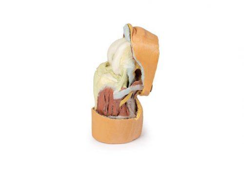 3D ANATOMY - Deep dissection of the knee joint, flexed
See details →
-
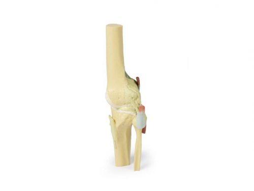 3D ANATOMY - Knee joint, extended
See details →
-
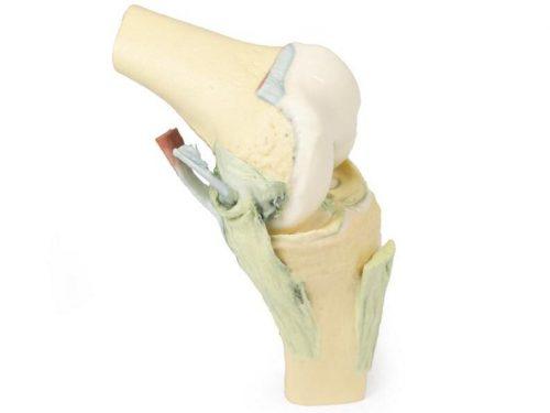 3D ANATOMY - Knee joint, flexed
See details →
-
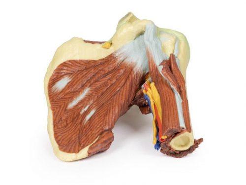 3D ANATOMY - Left shoulder, deep dissection of the joint, musculature, nerves and vessels
See details →
-
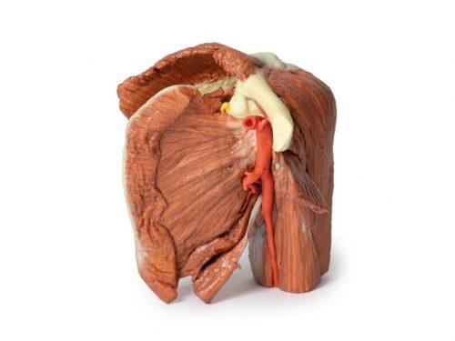 3D ANATOMY - Left shoulder, superficial muscles and axillary/brachial artery
See details →
-
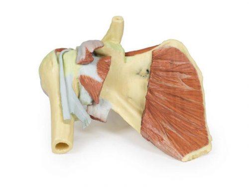 3D ANATOMY - Right shoulder, deep dissection of shoulder girdle, lateral clavicle and humerus
See details →
Is this equipment going into a new centre?
If you're equipping a classroom or a whole centre, starting from the shopping list works out expensive. Start from the needs assessment.