

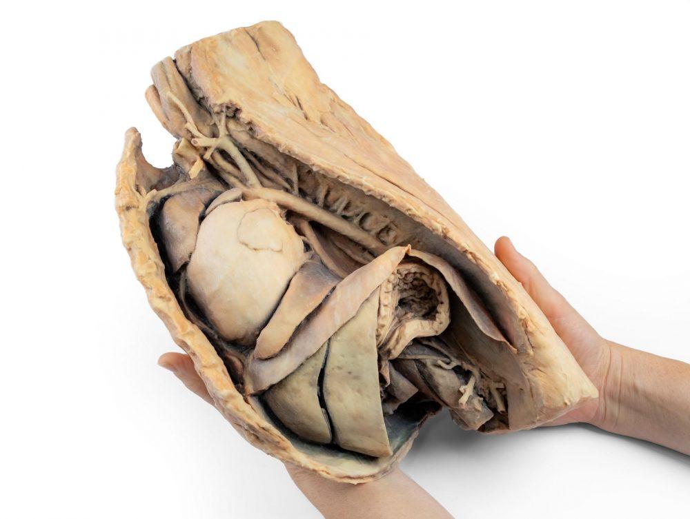
Reference 14446 · Anatomical model
Canine thoracic cavity left side
- Anatomical model
- Anatomy
- Main product
- Veterinary medicine
Left-side dissection of a dog’s thorax and cranial abdomen, with the left lung removed, showing the topography of the heart.
Price on request
Available in Spain, Portugal and Andorra · delivery time given in the proposal
Downloadable data sheet — features, specifications and delivery time, in PDF.
Need a technical report? Ask us for it and we'll send it within 24 h →
Description
What this equipment is and what it's designed for.
This specimen depicts the topography of the heart following removal of the left lung during a dissection of the thoracic cavity and cranial abdomen of a dog approached from the left side. The cranial and accessory lobes of the right lung remain in situ, serving as anatomical reference points. Within the cranial mediastinum, the brachiocephalic trunk and the left subclavian artery are identifiable as major branches arising from the aortic arch.
The thoracic portion of the oesophagus runs through the mediastinum in a craniocaudal direction. The diaphragm has been preserved to serve as a landmark for retrodiaphragmatic organs such as the liver and the stomach.
The diaphragmatic crura and their relationship to the aortic hiatus are maintained, as are the initial visceral branches of the abdominal aorta, specifically the coeliac trunk and the cranial mesenteric artery.
Both the interventricular and coronary grooves are visible. A portion of the right atrial wall has been removed to expose the right atrioventricular (tricuspid) valve, including the chordae tendineae and papillary muscles.
Additionally, the septomarginal trabecula (moderator band) is visible within the right ventricle, as well as the pathway from the ventricle to the pulmonary trunk. The left atrial wall is also open to display its lumen.
What you learn
Anatomical model modality. The competencies this equipment trains, in the language of the training plan.
- Identify the topography of the heart in the canine thorax
- Recognise the branches of the aortic arch
- Relate the diaphragm to the cranial abdominal organs
Cases and pathologies
The clinical scenarios that can be simulated with this equipment.
- Diaphragmatic hernia
- Persistent right fourth aortic arch
- Cardiomegaly
Features and what's included
What the equipment does and what comes in the box, as described by the manufacturer.
Features
- Left-side dissection of the thorax and cranial abdomen
- Cranial and accessory lobes of the right lung in situ
- Brachiocephalic trunk, left subclavian artery and thoracic oesophagus
- Diaphragm with aortic hiatus, coeliac trunk and cranial mesenteric artery
- Opened atria with visible tricuspid valve
Services for this equipment
Services matter: the equipment gets commissioned, the staff know how to use it and maintenance is settled before you start. Include them in your budget and avoid having to raise a second purchase.
- Preventive maintenance We'll let you know when it's due
Documentation
You can download it now or we'll send it on request.
Other related equipment
They share a specialty with this one. They train different things: none of them replaces it.
Is this equipment going into a new centre?
If you're equipping a classroom or a whole centre, starting from the shopping list works out expensive. Start from the needs assessment.