

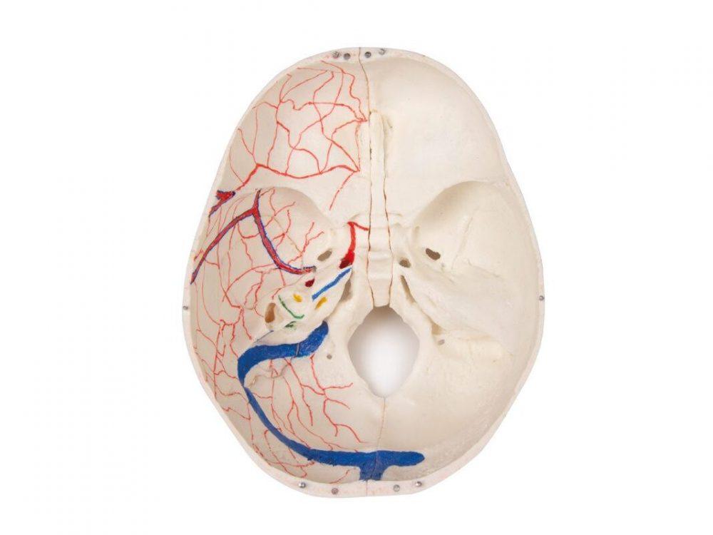
Reference 10992 · Anatomical model
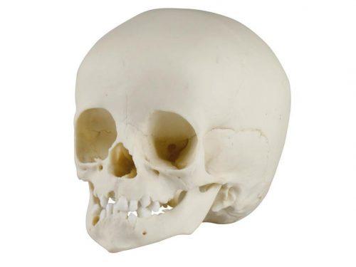
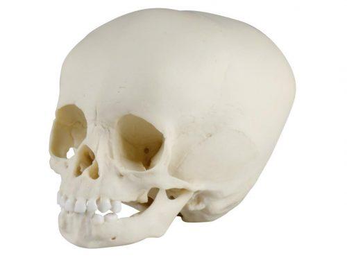
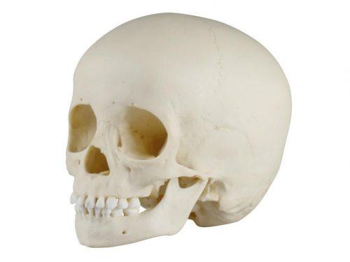
Luxury demonstration Children skull; 14 pieces; for advanced studies
- Anatomical model
- Anatomy
- Main product
- Medicine
This skull model is a cast of a real human children’s skull specimen and shows all anatomical structures in great detail. It was developed for students of anatomy, medicine, surgery, ENT medicine, ophthalmology and dentistry. The skull is complexly cut and joined together with metal and magnetic connections. The top of
Price on request
Available in Spain, Portugal and Andorra · delivery time given in the proposal
Downloadable data sheet — features, specifications and delivery time, in PDF.
Need a technical report? Ask us for it and we'll send it within 24 h →
Description
What this equipment is and what it's designed for.
This skull model is the cast of a real anatomical specimen of a human child’s skull and shows all the anatomical structures in great detail. It has been developed for students of anatomy, medicine, surgery, ENT, ophthalmology and dentistry. The skull has complex cuts and is joined by means of metal and magnetic connections.
The cranial vault is open and can be removed. The bony impressions of the sagittal sinus, of the transverse sinus and of the sigmoid sinus are painted, as are the meninges. The base of the skull is cut sagittally so that the cut passes through one cribriform plate on one side and another cut in the same plane through the other cribriform plate of the ethmoid, which leaves the crista galli and the perpendicular plate intact, as well as the whole nasal septum. The structures of the anterior, middle and posterior fossae are easily accessible. The nasal cavity, the turbinates, the septum and the bony pharynx and nasopharynx can be seen directly. The nasal septum can be separated from the surrounding bony structures. The removable temporal bone shows a complete external auditory canal.
The maxilla and the mandible show the structures of the teeth, the roots, the bony margin of the alveolar process and the dental nerves and vessels. The maxillary sinus can be opened by removing a flap. The teeth, including the permanent teeth still housed in the bone, become accessible by removing a bone flap.
Estimated age: 3 years
What you learn
Anatomical model modality. The competencies this equipment trains, in the language of the training plan.
- Identificar las estructuras del cráneo infantil en las tres fosas craneales.
- Reconocer la dentición definitiva alojada aún en el hueso.
- Localizar el tabique nasal, los cornetes y la nasofaringe.
- Describir las impresiones de los senos venosos y de las meninges.
Cases and pathologies
The clinical scenarios that can be simulated with this equipment.
- Docencia de anatomía pediátrica avanzada.
- Formación en odontopediatría y desarrollo de la dentición.
- Clases de otorrinolaringología y oftalmología infantil.
- Estudio de las vías de abordaje quirúrgico en el cráneo infantil.
Features and what's included
What the equipment does and what comes in the box, as described by the manufacturer.
Features
- Molde de una pieza anatómica real de cráneo infantil, en 14 partes.
- Cortes complejos unidos con conexiones metálicas y magnéticas.
- Bóveda craneal abierta y extraíble; hueso temporal extraíble con conducto auditivo externo completo.
- Maxilar y mandíbula con raíces, vasos y nervios dentales; seno maxilar abrible.
- Edad estimada de la pieza: 3 años.
Services for this equipment
Services matter: the equipment gets commissioned, the staff know how to use it and maintenance is settled before you start. Include them in your budget and avoid having to raise a second purchase.
- Preventive maintenance We'll let you know when it's due
Documentation
You can download it now or we'll send it on request.
Other related equipment
They share a specialty with this one. They train different things: none of them replaces it.
Is this equipment going into a new centre?
If you're equipping a classroom or a whole centre, starting from the shopping list works out expensive. Start from the needs assessment.