

Reference 11246 · Anatomical model
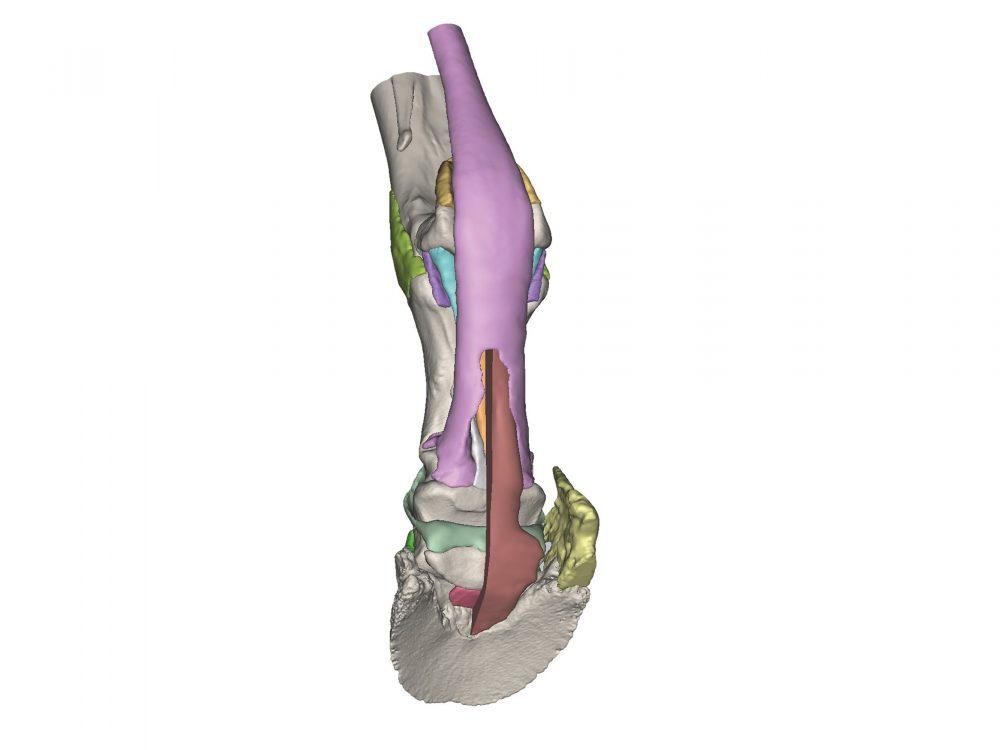
Foot of a horse as model - Model 3
- Anatomical model
- Anatomy
- Main product
- Veterinary medicine
Derived from CT and MR co-registered data, anatomically accurate and lifesize. 3D printed in full color, each anatomical component is individually colored. Hoof capsule is available separately and anchors the distal limb in the stance position. The series consists of four models: starting from Model 1 showing the full
Price on request
Available in Spain, Portugal and Andorra · delivery time given in the proposal
Downloadable data sheet — features, specifications and delivery time, in PDF.
Need a technical report? Ask us for it and we'll send it within 24 h →
Description
What this equipment is and what it's designed for.
Obtained from co-registered CT and MRI data, anatomically accurate and life size. 3D printed in full colour, with each anatomical component coloured separately. The hoof capsule is available separately and fixes the distal limb in the weight-bearing position. The series consists of four models: from model 1, which shows the complete anatomy of the foot with 25 anatomical structures, to progressively reduced models that show the deeper structures.
Model 3:
Similar to model 1, but with the deep digital flexor tendon sectioned, which shows its relationship with the distal sesamoid bone and the impar ligament. One of the ungular cartilages is missing in order to show the collateral ligaments of the distal sesamoid bone.
What you learn
Anatomical model modality. The competencies this equipment trains, in the language of the training plan.
- Identificar las estructuras profundas del pie del caballo.
- Relacionar el tendón flexor digital profundo con el hueso sesamoideo distal y el ligamento impar.
- Localizar los ligamentos colaterales del hueso sesamoideo distal.
- Relacionar la anatomía real con la imagen de CT y de MRI.
Cases and pathologies
The clinical scenarios that can be simulated with this equipment.
- Docencia de anatomía del pie equino.
- Patología del aparato podotroclear.
- Explicación de lesiones tendinosas al propietario.
Features and what's included
What the equipment does and what comes in the box, as described by the manufacturer.
Features
- Obtenido de datos corregistrados de CT y MRI, anatómicamente preciso y a tamaño natural.
- Impreso en 3D a todo color, con cada componente anatómico coloreado por separado.
- Modelo 3: tendón flexor digital profundo seccionado y ausencia de un cartílago ungular para dejar a la vista los ligamentos colaterales.
- Pertenece a una serie de cuatro modelos de detalle creciente.
Services for this equipment
Services matter: the equipment gets commissioned, the staff know how to use it and maintenance is settled before you start. Include them in your budget and avoid having to raise a second purchase.
- Preventive maintenance We'll let you know when it's due
Documentation
You can download it now or we'll send it on request.
Other related equipment
They share a specialty with this one. They train different things: none of them replaces it.
-
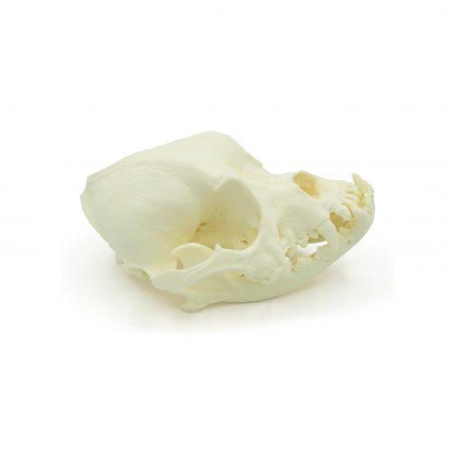 Brachycephalic canine skull - Anatomical
See details →
-
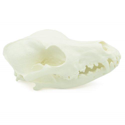 Complete canine skull - Anatomical
See details →
-
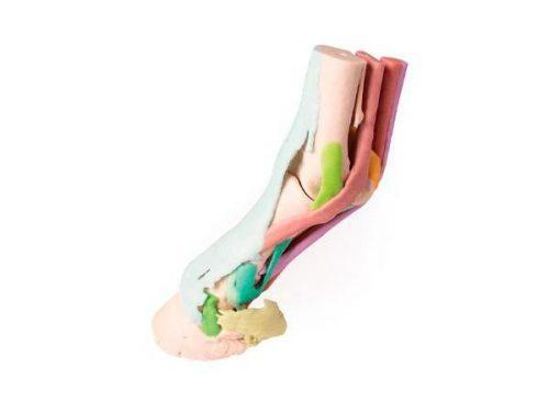 3D horse lower leg model - Educational - Model 1
See details →
- Anatomical model of cow front leg - 1/3 size See details →
-
 Bat skull model - Breed chiroptera sp.
See details →
-
 Canine femur - Anatomical
See details →
Is this equipment going into a new centre?
If you're equipping a classroom or a whole centre, starting from the shopping list works out expensive. Start from the needs assessment.