

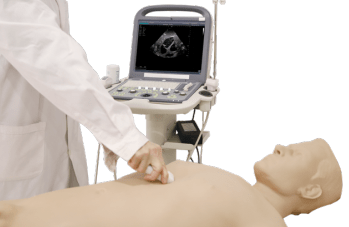
Reference 11689 · Task trainer
FAST Ultrasound Training Phantom
- Task trainer
- Emergencies
- Main product
- Medicine
This adult male torso (head to mid-thigh) is a realistic and anatomically correct phantom excellent for training users to develop and practice the skills necessary to perform FAST exam ultrasound procedures.
Price on request
Available in Spain, Portugal and Andorra · delivery time given in the proposal
Downloadable data sheet — features, specifications and delivery time, in PDF.
Need a technical report? Ask us for it and we'll send it within 24 h →
Description
What this equipment is and what it's designed for.
Outline
This adult male torso (head to mid-thigh) is a realistic and anatomically correct phantom excellent for training users to develop and practice the skills necessary to perform FAST exam ultrasound procedures.
Skills Gained
- Familiarity with chest, abdomen & pelvis underlying anatomy
- Ultrasound image interpretation
- Recognition of various pathologies using ultrasound
- Handling ultrasound machine & different transducers
- Palpation & various centeses
Features
- Anatomy:
- Lifelike feel and touch with soft abdomen.
- Internal simulated human skeleton structures like ribs, sternum, pelvis, etc.
- Correct chest & abdomen anatomical structures like heart, lungs, liver, gallbladder, pancreas, stomach, spleen, kidneys, intestinal canals, bladder, etc.
- Realism:
- The model is made of high polymer material which can be used with real clinical ultrasound machines of various types and brands.
- Real anatomical and pathological ultrasound images.
- Versatility: Bleeding in the thoracic, abdominal and pelvic cavity can be simulated. When internal bleeding happens, ultrasound check will show no echo liquid dark areas and simulated blood can be drawn.
- Key Features:
- Chest ultrasound functions
A.The product can simulate pleural effusion, such as pericardial effusion and show related costophrenic angle as well as liquid dark area. The amount of pleural effusion can be changed by adjusting injected fluid volume.
B.Ultrasound anatomical structures include heart with left ventricle, right ventricle, left atrium, right atrium, cardiac valves, etc.
C.Standard sub-xiphoid four-chamber cardiac view is available. Support separate display of ventricle, atrium, mitral valve, tricuspid valve, aortic valve, pulmonary valve, pulmonary artery, aorta and other structures.
D.Realistic right lung structure allows for using the probe long-axis to scan along the parasternal line, anterior axillary line, mid-axillary, posterior axillary line and the outer edge line of scapula. Users can also do short-axis scanning for ultrasound images of organs, pleural line, ribs and sternum.
- Abdominal ultrasound functions
A. Bony landmarks include costal margin, xiphoid, anterior superior iliac spine, etc.
B. Scan the hepatorenal space and splenorenal space to check if ascites exits. No echo if there is abdominal effusion. The amount of ascites can be changed by adjusting injected fluid volume.
C. Various pathologies and anatomical ultrasound images available, like Glisson’s capsule, liver parenchyma, hepatic veins, pancreatic parenchyma, pancreatic duct, splenic capsule, parenchyma of the spleen, renal capsule, renal cortex, renal medullary, renal pelvis, renal calyx, etc.
D. A full bladder in the abdominal cavity will show pelvic ultrasound features as well as liquid dark area of pelvic effusion.
Specifications
- Gross weight (kg): 59
- Packing size (cm): 116 x 57 x 36
What you learn
Task trainer modality. The competencies this equipment trains, in the language of the training plan.
- Reconocer la anatomía del tórax, el abdomen y la pelvis con la sonda.
- Realizar la secuencia completa de la ecografía FAST.
- Interpretar la imagen ecográfica normal y patológica.
- Manejar las distintas sondas del ecógrafo.
- Localizar el punto de punción y aspirar líquido.
Related procedures: Abdominal · Ultrasound
Cases and pathologies
The clinical scenarios that can be simulated with this equipment.
- Hemorragia interna torácica, abdominal y pélvica.
- Derrame pleural y derrame pericárdico.
- Ascitis y derrame pélvico.
- Politraumatizado en la valoración inicial.
Features and what's included
What the equipment does and what comes in the box, as described by the manufacturer.
Features
- Torso masculino de la cabeza a la mitad del muslo, con abdomen blando.
- Estructuras óseas internas simuladas: costillas, esternón y pelvis.
- Órganos torácicos y abdominales completos: corazón, pulmones, hígado, vesícula, páncreas, estómago, bazo, riñones, asas y vejiga.
- Material polimérico compatible con ecógrafos clínicos reales de distintas marcas.
- La cantidad de derrame se modifica ajustando el volumen de líquido inyectado.
Services for this equipment
Services matter: the equipment gets commissioned, the staff know how to use it and maintenance is settled before you start. Include them in your budget and avoid having to raise a second purchase.
- Preventive maintenance We'll let you know when it's due
Documentation
You can download it now or we'll send it on request.
Other related equipment
They train the same technique. Each one approaches it in its own way: none of them replaces this one.
-
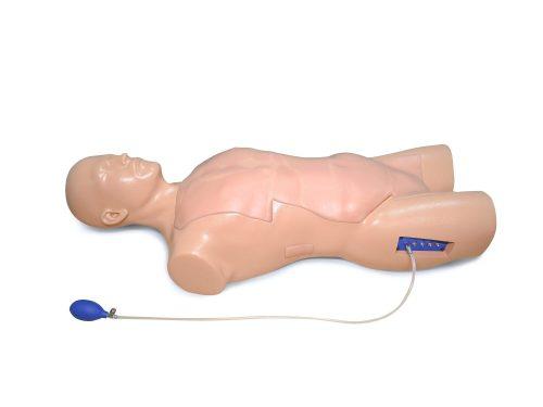 Adult torso simulator for FAST and eFAST ultrasound - TTE probe
See details →
-
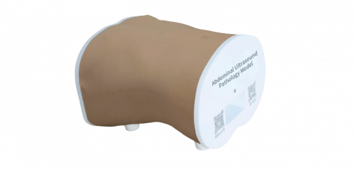 Abdominal Ultrasound Pathology Model
See details →
-
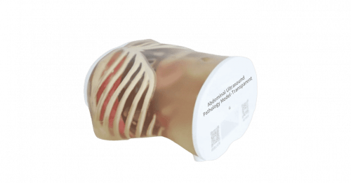 Abdominal Ultrasound Pathology Model-Transparent
See details →
-
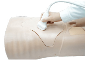 Abdominal Ultrasound Phantom
See details →
-
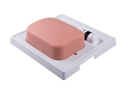 Gallblader model for ultrasound - SONOTRAIN
See details →
-
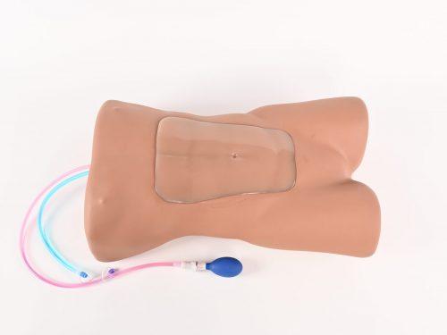 Simulator for abdominal aortic aneurysm ultrasound assessment
See details →
Is this equipment going into a new centre?
If you're equipping a classroom or a whole centre, starting from the shopping list works out expensive. Start from the needs assessment.