

Reference 03995 · Anatomical model
Eye in orbit model, Magnification x5 - 11 Parts
- Anatomical model
- Anatomy
- Main product
- Medicine
A 5x-magnified model of the eye within its bony orbit, with the eyeball dissected into two halves and several removable parts.
Price on request
Available in Spain, Portugal and Andorra · delivery time given in the proposal
Downloadable data sheet — features, specifications and delivery time, in PDF.
Need a technical report? Ask us for it and we'll send it within 24 h →
Description
What this equipment is and what it's designed for.
Magnified 5 times life size, this model shows the eyeball with the optic nerves and muscles in their natural position within the bony orbit. The eyeball is dissected into 2 removable halves, with the internal structures fully exposed.
The removable parts are: cornea, iris, lens and vitreous body (4 parts); the 3 rectus bulbi muscles; the levator palpebrae superioris muscle together with the lacrimal gland; the eyeball with extraocular muscles (2 parts); and the base of the bony orbit.
Mounted on base. Size: 40x26x31.5 cm. Weight: approx. 1300 g.
What you learn
Anatomical model modality. The competencies this equipment trains, in the language of the training plan.
- Identificar sobre el modelo las estructuras del globo ocular y su órbita.
- Reconocer la córnea, el iris, el cristalino, la retina, el nervio óptico y la musculatura extraocular.
- Relacionar la forma de cada estructura con la función que desempeña.
- Describir la región empleando la terminología anatómica correcta.
- Orientar la pieza según los planos y ejes anatómicos.
Cases and pathologies
The clinical scenarios that can be simulated with this equipment.
- Explicar la acomodación, la miopía, la hipermetropía y la presbicia.
- Relacionar la presión intraocular con el glaucoma y la retina con el desprendimiento.
- Apoyar la exploración de la motilidad ocular y del fondo de ojo.
Features and what's included
What the equipment does and what comes in the box, as described by the manufacturer.
Features
- Reproduce la región en tres dimensiones: se puede rotar y observar desde cualquier ángulo.
- Se desmonta en piezas separables para acceder a las estructuras internas.
- Reproduce la estructura a escala ampliada, de modo que los detalles se ven a simple vista.
- Sirve tanto para la explicación ante el grupo como para el estudio individual del alumno.
- Pieza pensada para manipulación repetida en aula y laboratorio, sin consumibles asociados.
Includes
- Mounting base
Services for this equipment
Services matter: the equipment gets commissioned, the staff know how to use it and maintenance is settled before you start. Include them in your budget and avoid having to raise a second purchase.
- Preventive maintenance We'll let you know when it's due
Documentation
You can download it now or we'll send it on request.
Other related equipment
They share a specialty with this one. They train different things: none of them replaces it.
-
 3D ANATOMY - Superior orbit section
See details →
-
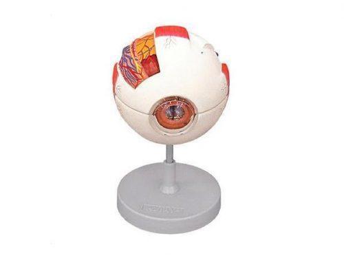 Anatomical eye model - Life size x6 - 6 parts
See details →
-
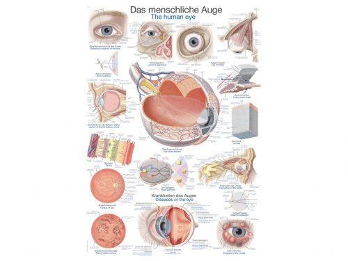 Chart The human eye, 50x70cm
See details →
-
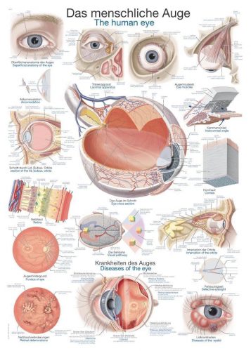 Chart The human eye, 70x100cm
See details →
-
 Eye model - 4 times life size - 6 parts
See details →
-
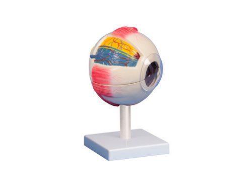 Eye model - 6 times life size - 6 parts
See details →
Is this equipment going into a new centre?
If you're equipping a classroom or a whole centre, starting from the shopping list works out expensive. Start from the needs assessment.