

Reference 14441 · Anatomical model
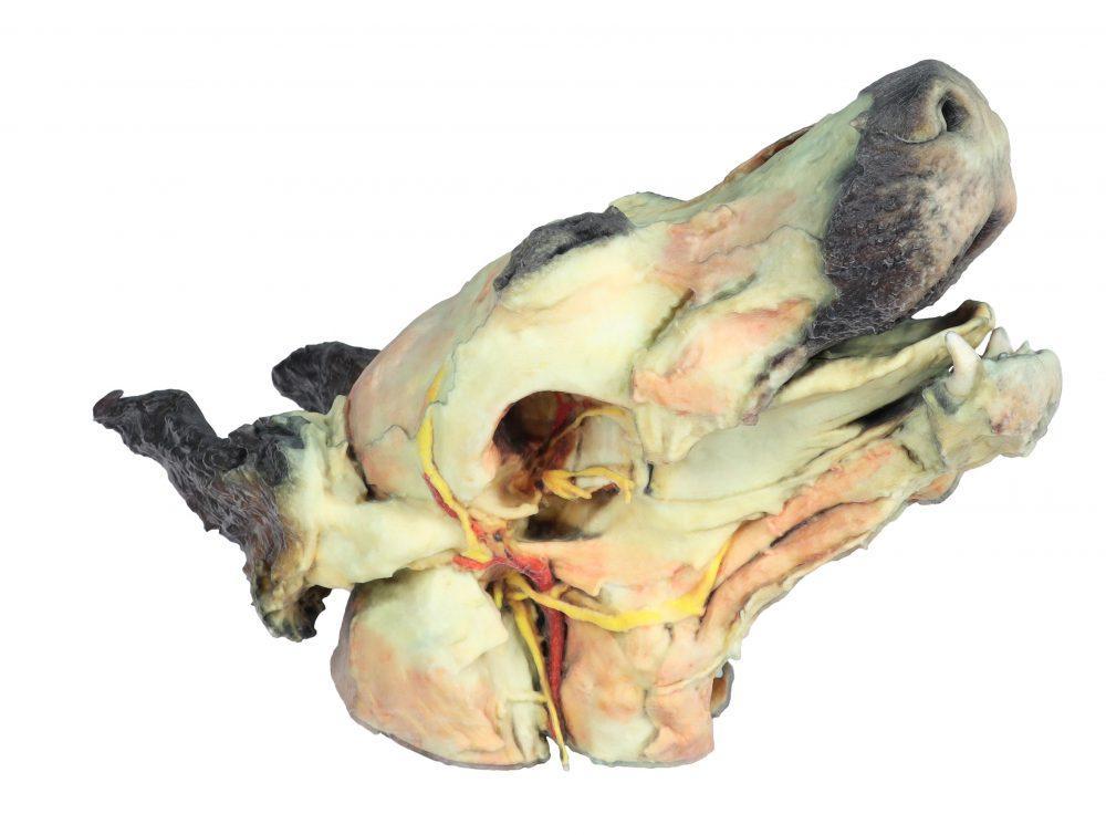
Dog head - superficial and deep dissections
- Anatomical model
- Anatomy
- Main product
- Veterinary medicine
Dog head with a superficial dissection on the left side and a deep dissection, with the mandible removed, on the right side.
Price on request
Available in Spain, Portugal and Andorra · delivery time given in the proposal
Downloadable data sheet — features, specifications and delivery time, in PDF.
Need a technical report? Ask us for it and we'll send it within 24 h →
Description
What this equipment is and what it's designed for.
Superficial anatomical structures:
- Tip of the nose (Apex nasi)
- Right and left wings of the nose (Alae nasi)
- Nonglandular skin of the tip of the nose (Planum nasale)
Superficial dissection on the left side:
On the left side, the skin has been removed to identify the main anatomical structures, which are described in groups below.
Muscles of the facial neuromuscular system:
- Levator nasolabialis muscle (M. levator nasolabialis)
- Caninus muscle (M. caninus)
- Buccinator muscle (M. buccinator)
- Zygomaticus muscle (M. zygomaticus)
- Parotidoauricularis muscle (M. parotidoauricularis)
Muscles of the mandibular neuromuscular system (masticatory muscles):
- Masseter muscle (M. masseter)
- Temporal muscle (M. temporalis)
Nerves:
- Facial nerve (N. facialis)
- Dorsal buccal branch (Rami buccales)
- Ventral buccal branch (Rami buccales)
- Buccolabial branches (Rami buccolabiales)
- Auriculopalpebral nerve (N. auriculopalpebralis)
Vessels:
- Facial artery (A. facialis)
- External jugular vein (V. jugularis externa), with the maxillary vein (V. maxillaris) and the linguofacial vein (V. linguofacialis)
Salivary glands:
- Parotid gland and parotid duct (Glandula parotis, Ductus parotideus)
- Mandibular gland (Glandula mandibularis)
Lymph nodes:
- Mandibular lymph nodes (Lnn. mandibulares)
Deep dissection on the right side:
On the right side the mandible has been removed to show deeper anatomical structures. The surfaces of the temporal bone for the temporomandibular joint (Articulatio temporomandibularis) can be seen. The medial pterygoid muscle (M. pterygoideus medialis) is identified and sectioned at its insertion on the ramus of the mandible (Ramus mandibulae). Next to this muscle, the branches of the mandibular nerve (N. mandibularis) are identified, as well as the maxillary nerve (N. maxillaris) and the maxillary artery (A. maxillaris). Near the external acoustic meatus (Meatus acusticus externus), the facial nerve (N. facialis) has been preserved with one of its branches, the auriculopalpebral nerve (N. auriculopalpebralis), which runs parallel to the zygomatic arch (Arcus zygomaticus) and is visible after removal of the parotid salivary gland (Glandula parotis). The tongue is shown in its entire caudal extent, reached by the styloglossus (M. styloglossus), genioglossus (M. genioglossus) and geniohyoid (M. geniohyoideus) muscles. Next to these muscles, the hypoglossal nerve (N. hypoglossus) is identified. In relation to the pharynx (Pharynx), the pharyngeal constrictor muscles (Mm. constrictores pharyngis caudales) are identified. In the most caudal area, in relation to the neck, the course of the common carotid artery (A. carotis communis) and the vagosympathetic trunk (Truncus vagosympathicus) are identified.
What you learn
Anatomical model modality. The competencies this equipment trains, in the language of the training plan.
- Identify the facial and masticatory musculature of the dog
- Follow the course of the facial nerve and its branches
- Recognise the salivary glands and lymph nodes of the head
- Identify the deep structures of the temporomandibular joint and the tongue
Cases and pathologies
The clinical scenarios that can be simulated with this equipment.
- Facial nerve paralysis
- Salivary mucocele
- Enlarged mandibular lymph nodes
Features and what's included
What the equipment does and what comes in the box, as described by the manufacturer.
Features
- Superficial dissection of the left side
- Deep dissection of the right side, with the mandible removed
- Facial and masticatory muscles, nerves, vessels, salivary glands and lymph nodes
- Latin anatomical nomenclature in the description
Services for this equipment
Services matter: the equipment gets commissioned, the staff know how to use it and maintenance is settled before you start. Include them in your budget and avoid having to raise a second purchase.
- Preventive maintenance We'll let you know when it's due
Documentation
You can download it now or we'll send it on request.
Other related equipment
They share a specialty with this one. They train different things: none of them replaces it.
-
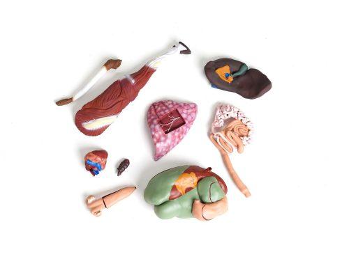 Bovine model, 15 pieces, 1/3 natural size
See details →
-
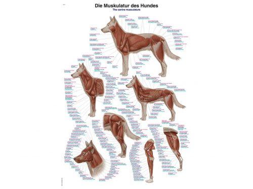 Canine musculature chart, 50x70cm, 200g - German, Latin and English
See details →
-
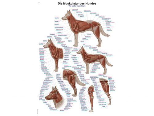 Chart The canine musculature, 70x100cm
See details →
-
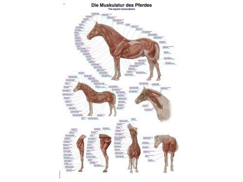 Chart The equine musculature, 70x100cm
See details →
-
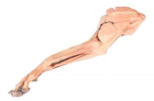 Dog forelimb - muscles, tendons, ligaments, vessels and nerves distal to the elbow
See details →
-
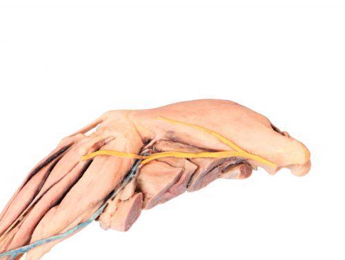 Dog hindlimb - muscles, tendons, ligaments, vessels and nerves distal to the stifle
See details →
Is this equipment going into a new centre?
If you're equipping a classroom or a whole centre, starting from the shopping list works out expensive. Start from the needs assessment.