

Reference 02718 · Anatomical model
3D ANATOMY - Superficial and deep structures of the distal leg and foot
- Anatomical model
- Anatomy
- Main product
- Medicine
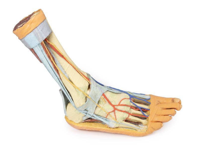
Superficial and deep structures of the distal leg and foot in a single specimen.
Price on request
Available in Spain, Portugal and Andorra · delivery time given in the proposal
Downloadable data sheet — features, specifications and delivery time, in PDF.
Need a technical report? Ask us for it and we'll send it within 24 h →
Description
What this equipment is and what it's designed for.
This 3D printed anatomical specimen showcases both superficial and deep structures of the distal leg and foot.
Posterior Compartment: Deep Musculature and Neurovascular Structures
The posterior compartment of the leg has been dissected proximally to remove the triceps surae muscles and the tendocalcaneus, revealing the deep muscles: tibialis posterior, flexor digitorum longus, and flexor hallucis longus. The tibial nerve and posterior tibial artery can be traced down to their medial and lateral plantar branches at the level of the flexor retinaculum. To enhance visibility, the origin of the abductor hallucis brevis has been removed.
Venous Anatomy and Anterior Compartment Highlights
The great saphenous vein is preserved as it originates from the medial side of the dorsal venous arch, extending to the edge of the specimen. While the anterior compartment muscles have been removed to expose the interosseous membrane, the anterior tibial artery, and the deep fibular nerve, key tendons remain intact. These include the insertions of tibialis anterior, extensor hallucis longus, and the hallucal tendon of extensor digitorum longus, all passing deep to the inferior extensor retinaculum. The anterior tibial artery continues as dorsalis pedis, giving rise to the arcuate artery and dorsal metatarsal arteries. Removal of the dorsal interossei allows visualization of their terminal branches as they approach the plantar interossei.
Lateral View: Muscles, Tendons, and Nerve Terminations
On the lateral aspect, the fibularis longus and fibularis brevis muscles and their tendons are clearly visible. The tendons pass beneath the superior fibular retinaculum (cut edge) and the intact inferior fibular retinaculum. Also visible are the extensor digitorum longus tendon to the fifth digit and the termination of the superficial fibular nerve. Near the entry of the fibularis longus tendon into the plantar foot, the abductor digiti minimi muscle origin is preserved.
Ligamentous Structures
Deep structures include key ligaments of the distal leg and foot, such as the anterior and posterior tibiofibular ligaments, calcaneofibular ligament, dorsal and posterior talonavicular ligaments, and the deltoid ligament, all providing essential support and insight into joint stability.
What you learn
Anatomical model modality. The competencies this equipment trains, in the language of the training plan.
- Compare the superficial and the deep plane in the distal leg and foot.
- Identify the tendons, vessels and nerves crossing the ankle.
- Place the palpable landmarks of the ankle for examination and puncture.
Cases and pathologies
The clinical scenarios that can be simulated with this equipment.
- Ankle sprain and fracture.
- Tarsal tunnel syndrome.
- Distal ischaemia and pulse assessment.
- Ankle block.
Features and what's included
What the equipment does and what comes in the box, as described by the manufacturer.
Features
- 3D printed replica made from radiological data or from a dissected cadaveric specimen, not a stylised model: this is real anatomy.
- Developed and checked by the anatomists of the Human Anatomy Teaching Centre at Monash University.
- Colourised to distinguish muscle, nerve, artery, vein and bone, and to separate territories or segments.
- Not human tissue: no restrictions on transport, import or use in institutions without permission to work with cadavers.
- Avoids the ethical and preservation problems that come with plastination.
- Supplied with a detailed description of the anatomy represented in the specimen.
Services for this equipment
Services matter: the equipment gets commissioned, the staff know how to use it and maintenance is settled before you start. Include them in your budget and avoid having to raise a second purchase.
- Preventive maintenance We'll let you know when it's due
Documentation
You can download it now or we'll send it on request.
Other related equipment
They share a specialty with this one. They train different things: none of them replaces it.
Is this equipment going into a new centre?
If you're equipping a classroom or a whole centre, starting from the shopping list works out expensive. Start from the needs assessment.