

Reference 02679 · Anatomical model
3D ANATOMY - Posterior abdominal wall
- Anatomical model
- Anatomy
- Main product
- Medicine
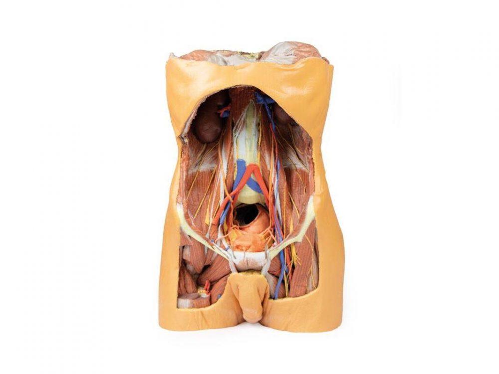
Male posterior abdominal wall from the diaphragm to the pelvic brim, including pelvis and proximal thigh.
Price on request
Available in Spain, Portugal and Andorra · delivery time given in the proposal
Downloadable data sheet — features, specifications and delivery time, in PDF.
Need a technical report? Ask us for it and we'll send it within 24 h →
Description
What this equipment is and what it's designed for.
This detailed 3D printed model presents the male posterior abdominal wall from the diaphragm down to the pelvic brim, including the pelvis and proximal thigh.
A focused pelvic and thigh version is also available (MP1770).
Muscular Anatomy & Diaphragm
The parietal peritoneum is removed to expose the key muscles of the posterior abdominal wall: psoas major, quadratus lumborum, transversus abdominis, and iliacus below the iliac crest. The diaphragm shows distinct muscular fibers originating from the thoracic cage and lumbar vertebrae (right crus L1–L3, left crus L1–L2), connected by the median arcuate ligament. Major diaphragmatic openings for the esophagus, aorta, and inferior vena cava are visible, although the aorta is removed.
Nerves
Somatic nerves are clearly identifiable, including the subcostal, iliohypogastric, and ilioinguinal nerves (which arise together in this specimen), the lateral cutaneous nerve of the thigh, the genitofemoral nerve (on psoas), and the femoral nerve situated between psoas and iliacus. The sympathetic trunks run alongside the lumbar vertebrae.
Vessels & Kidneys
The aorta and inferior vena cava are transected at L3, with the aortic bifurcation positioned slightly higher than usual. The renal arteries and veins are preserved, though their full origin is partly obscured by the absence of the great vessels. Both kidneys are dissected free from surrounding fat, showing the typical lower position of the right kidney. The ureters are visible descending from the renal hilum, passing medial to the psoas before crossing the pelvic brim into the true pelvis.
This model offers an exceptional view of the complex anatomy of the posterior abdominal wall, pelvis, and proximal thigh, making it ideal for advanced anatomical study, surgical planning, and clinical reference.
What you learn
Anatomical model modality. The competencies this equipment trains, in the language of the training plan.
- Identify the muscles of the posterior abdominal wall and the diaphragm.
- Locate the kidney, the ureter and the great vessels in the retroperitoneum.
- Follow the nerves of the lumbar plexus between the muscle planes.
- Relate the anatomy to retroperitoneal approaches and to abdominal wall blocks.
Cases and pathologies
The clinical scenarios that can be simulated with this equipment.
- Renal and ureteric disease approached retroperitoneally.
- Psoas abscess.
- Abdominal aortic aneurysm.
- Lumbar plexus and quadratus lumborum blocks.
Features and what's included
What the equipment does and what comes in the box, as described by the manufacturer.
Features
- 3D printed replica made from radiological data or from a dissected cadaveric specimen, not a stylised model: this is real anatomy.
- Developed and checked by the anatomists of the Human Anatomy Teaching Centre at Monash University.
- Colourised to distinguish muscle, nerve, artery, vein and bone, and to separate territories or segments.
- Not human tissue: no restrictions on transport, import or use in institutions without permission to work with cadavers.
- Avoids the ethical and preservation problems that come with plastination.
- Supplied with a detailed description of the anatomy represented in the specimen.
Services for this equipment
Services matter: the equipment gets commissioned, the staff know how to use it and maintenance is settled before you start. Include them in your budget and avoid having to raise a second purchase.
- Preventive maintenance We'll let you know when it's due
Documentation
You can download it now or we'll send it on request.
Other related equipment
They share a specialty with this one. They train different things: none of them replaces it.
Is this equipment going into a new centre?
If you're equipping a classroom or a whole centre, starting from the shopping list works out expensive. Start from the needs assessment.