

Reference 02636 · Anatomical model
3D ANATOMY - Nervous system dissection
- Anatomical model
- Nervous system
- Main product
- Medicine
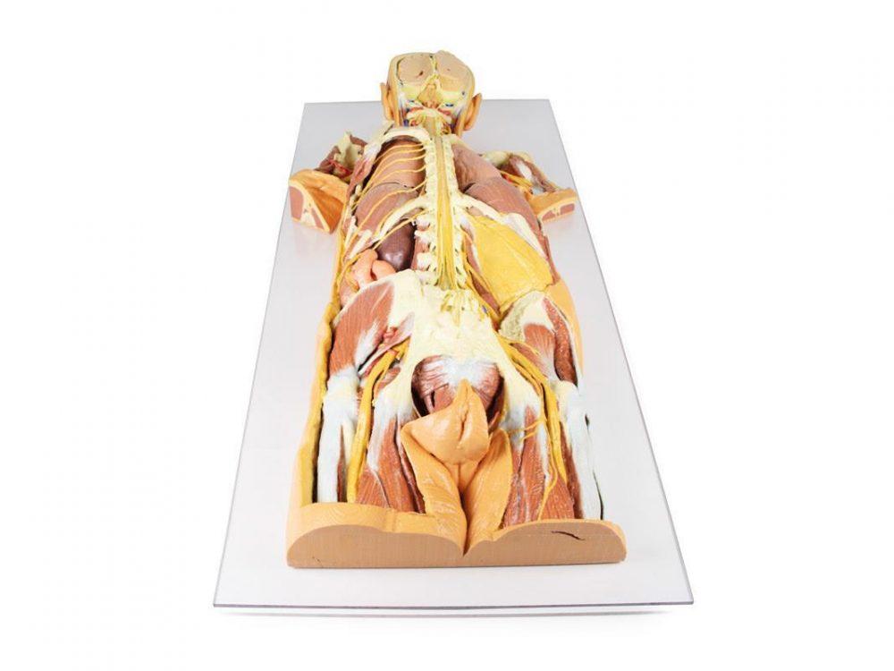
Dorsal view of head, neck, thorax, abdomen and gluteal regions, centred on the central nervous system and segmental nerves.
Price on request
Available in Spain, Portugal and Andorra · delivery time given in the proposal
Downloadable data sheet — features, specifications and delivery time, in PDF.
Need a technical report? Ask us for it and we'll send it within 24 h →
Description
What this equipment is and what it's designed for.
This detailed 3D printed specimen provides a unique dorsal view of the head, neck, thorax, abdomen, and gluteal regions, highlighting the central nervous system and segmental nerves in anatomical context.
The model reveals deep structures through extensive removal of posterior elements, including the posterior cranium, vertebral arches, and parts of the thoracic and gluteal walls.
Cranial and Cervical Structures
The posterior skull is removed to display a coronal section of the cerebral hemispheres, separated by the falx cerebri and bordered by a preserved superior sagittal sinus. The cerebellum is removed, exposing the fourth ventricle, pons, medulla oblongata, and cranial nerves VII–XII. The vertebral arteries are visible ascending through the transverse foramina and curving toward the foramen magnum. The cervical and brachial plexus roots are shown resting on scalene muscles and major vessels, extending into the axilla.
Axilla and Upper Thorax
The scapulae are partially (left) or fully (right) removed, allowing visualization of the brachial plexus and axillary vessels.
On the left, parts of deltoid, infraspinatus, and teres minor remain, while the long thoracic nerve, lateral thoracic artery, and subscapular branches are preserved.
On the right, the axillary nerve and posterior circumflex humeral artery are shown passing toward the surgical neck of the humerus.
Thoracic Spine and Intercostal Nerves
A laminectomy reveals the spinal cord, dorsal roots, and ganglia.
On the right, the posterior thoracic wall is removed (ribs 2–12), exposing the lung and diaphragm.
On the left, ribs 3–5 are retained to show intercostal musculature and nerve positioning, with the full set of intercostal nerves preserved.
Lumbar Region and Abdomen
The lumbar spinal cord is opened to show the conus medullaris and cauda equina.
On the right, the quadratus lumborum is removed, exposing subcostal and lumbar nerves along the psoas.
On the left, the subcostal nerve, kidney, and jejunum are visible through an opened peritoneal cavity.
Gluteal and Pelvic Dissection
In the gluteal region, gluteus maximus is removed bilaterally:
Left side: Gluteus medius is preserved; the superior and inferior gluteal arteries and nerves, internal pudendal vessels, and the sciatic nerve are visible relative to the piriformis and lateral rotators.
Right side: Deeper dissection reveals the lumbar and sacral plexuses within the true pelvis.
This model offers a rare, continuous view of deep axial anatomy, perfect for advanced anatomical study, neuroanatomy training, and surgical reference.
What you learn
Anatomical model modality. The competencies this equipment trains, in the language of the training plan.
- Follow the continuity between spinal cord, roots, plexuses and peripheral nerves.
- Identify the cranial nerves at their origin.
- Relate the nerve course to the dermatomes and myotomes.
- Explain the deficit to be expected from the level of the lesion.
Cases and pathologies
The clinical scenarios that can be simulated with this equipment.
- Spinal cord injury and its functional level.
- Cervical and lumbar radiculopathy.
- Brachial plexus injuries.
- Peripheral nerve compression syndromes.
Features and what's included
What the equipment does and what comes in the box, as described by the manufacturer.
Features
- 3D printed replica made from radiological data or from a dissected cadaveric specimen, not a stylised model: this is real anatomy.
- Developed and checked by the anatomists of the Human Anatomy Teaching Centre at Monash University.
- Colourised to distinguish muscle, nerve, artery, vein and bone, and to separate territories or segments.
- Not human tissue: no restrictions on transport, import or use in institutions without permission to work with cadavers.
- Avoids the ethical and preservation problems that come with plastination.
- Supplied with a detailed description of the anatomy represented in the specimen.
Services for this equipment
Services matter: the equipment gets commissioned, the staff know how to use it and maintenance is settled before you start. Include them in your budget and avoid having to raise a second purchase.
- Preventive maintenance We'll let you know when it's due
Documentation
You can download it now or we'll send it on request.
Other related equipment
They share a specialty with this one. They train different things: none of them replaces it.
-
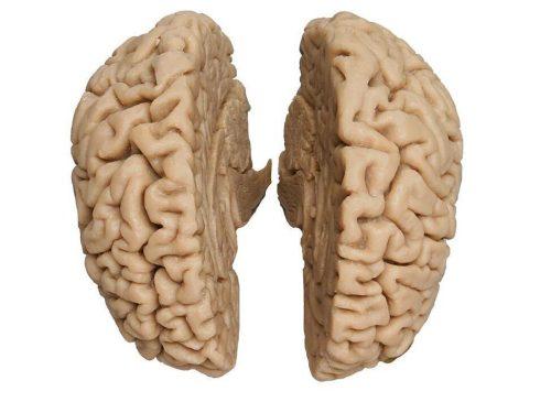 3D ANATOMY - 3D human brain
See details →
-
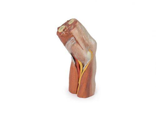 3D ANATOMY - Cubital fossa (muscles, major nerves and brachial artery)
See details →
-
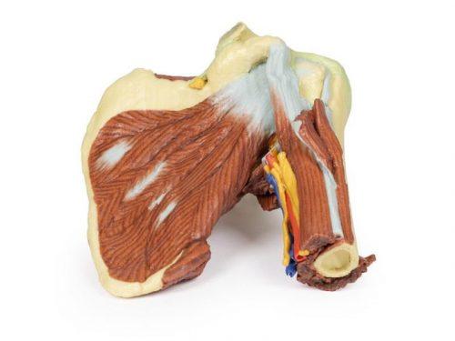 3D ANATOMY - Left shoulder, deep dissection of the joint, musculature, nerves and vessels
See details →
-
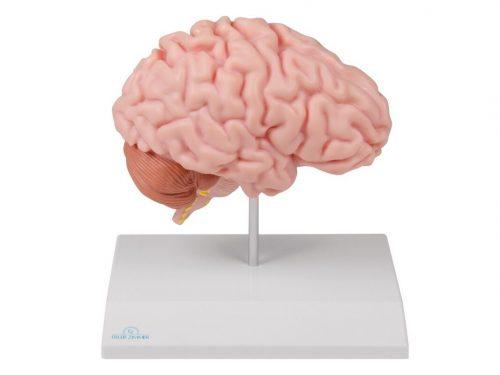 Anatomical brain half, life-size - EZ Augmented Anatomy
See details →
-
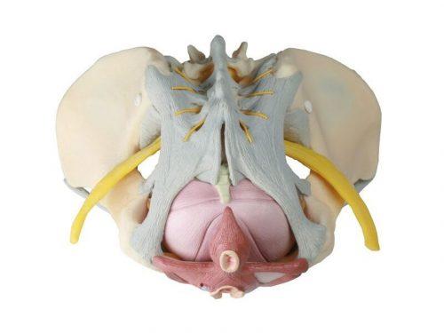 Anatomical model of female pelvis with ligaments, nerves and musculature
See details →
-
 Anatomical model of neuron, Magnification x2500 - 2 Parts
See details →
Is this equipment going into a new centre?
If you're equipping a classroom or a whole centre, starting from the shopping list works out expensive. Start from the needs assessment.