

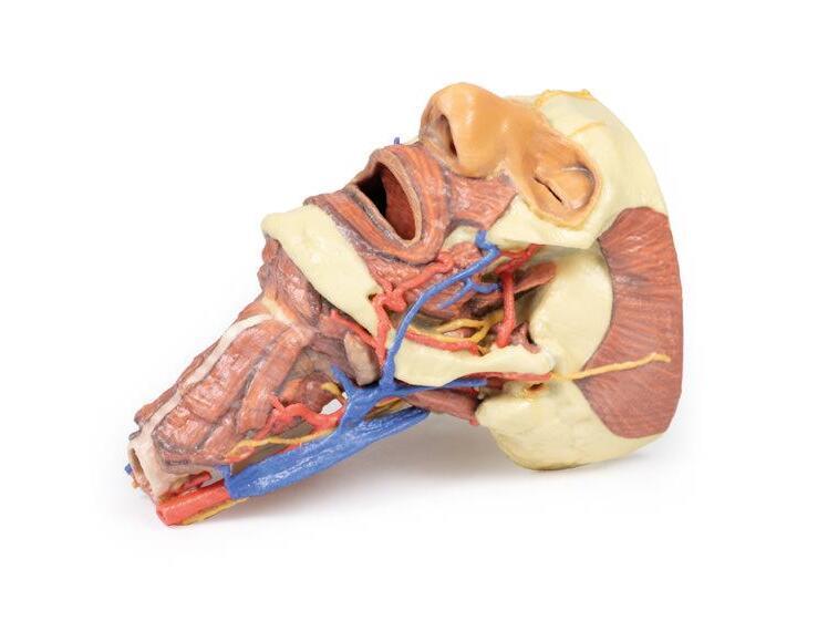
Reference 02677 · Anatomical model
3D ANATOMY - Head and visceral column of the neck
- Anatomical model
- Anatomy
- Main product
- Medicine
Head and visceral column of the neck: muscles, nerves, arteries, veins and cranial structures.
Price on request
Available in Spain, Portugal and Andorra · delivery time given in the proposal
Downloadable data sheet — features, specifications and delivery time, in PDF.
Need a technical report? Ask us for it and we'll send it within 24 h →
Description
What this equipment is and what it's designed for.
This 3D print provides a detailed exploration of the head and visceral column of the neck, showcasing muscles, nerves, arteries, veins, and cranial structures.
Facial Anatomy
The right side of the face is dissected to reveal the facial nerve and its branches (temporal, zygomatic, buccal, marginal mandibular, and cervical) following removal of the parotid gland. The relationships of the facial nerve, retromandibular vein, and external carotid artery are clearly visible. Muscles of the head include temporalis, masseter, and posterior belly of digastric, while arteries such as the facial, transverse facial, and superficial temporal arteries are preserved. The facial and transverse facial veins unite to form the common facial vein, which joins the retromandibular vein to form the external jugular vein.
From the anterior view, facial muscles around the mouth (buccinator, orbicularis oris, zygomaticus major) and muscles of mastication are displayed. In the left infratemporal fossa, the medial and lateral pterygoids are dissected to show the mandibular division of the trigeminal nerve, including the lingual nerve and inferior alveolar branch. The ophthalmic division branches (supraorbital and supratrochlear nerves) supplying the forehead and scalp are also demonstrated. The submandibular glands and facial vessels coursing over the mandible are clearly visible.
Neck Anatomy
The musculoskeletal structures of the neck are removed to expose the pharynx posteriorly, larynx anteriorly, and neurovascular bundles laterally. The suprahyoid and infrahyoid muscles are visible, as are the vocal folds viewed from below the trachea. The cricothyroid muscle is also identifiable. Neurovascular structures include the hypoglossal nerve, vagus nerve, internal jugular vein, common carotid artery with bifurcation into ECA and ICA, and the superior thyroid artery. The ansa cervicalis and internal branch of the superior laryngeal nerve are visible on the anterior neck.
Posterior Pharynx and Vertebrobasilar Structures
The superior, middle, and inferior constrictors of the pharynx are displayed, along with the esophagus and posterior horn of the hyoid bone. The carotid sheath shows the vagus nerve and its pharyngeal branches, and the recurrent laryngeal nerve is briefly visible. Occipital arteries curve around the mastoid, and the vertebral arteries are seen entering the foramen magnum. Removal of the cerebellum exposes the fourth ventricle, cut cerebellar peduncles, and the posterior inferior cerebellar artery on the right.
Cranial Cavity and Visual Pathways
The orbits are opened to reveal orbital nerves, vessels, eyes, and optic nerves. The optic chiasm, optic tracts, and lateral geniculate bodies are retained, showing key visual pathways. The brainstem is cut to demonstrate cranial nerve origins, while the olfactory tracts and bulbs are also displayed.
What you learn
Anatomical model modality. The competencies this equipment trains, in the language of the training plan.
- Identify the visceral column of the neck: larynx, trachea, pharynx, oesophagus and thyroid.
- Place its relations with the neurovascular bundle of the neck.
- Recognise the course of the recurrent laryngeal nerve.
- Relate the anatomy to tracheostomy and to thyroid surgery.
Cases and pathologies
The clinical scenarios that can be simulated with this equipment.
- Surgical thyroid disease.
- Recurrent nerve injury and postoperative dysphonia.
- Tracheostomy and stoma care.
- Tumours of the larynx and hypopharynx.
Features and what's included
What the equipment does and what comes in the box, as described by the manufacturer.
Features
- 3D printed replica made from radiological data or from a dissected cadaveric specimen, not a stylised model: this is real anatomy.
- Developed and checked by the anatomists of the Human Anatomy Teaching Centre at Monash University.
- Colourised to distinguish muscle, nerve, artery, vein and bone, and to separate territories or segments.
- Not human tissue: no restrictions on transport, import or use in institutions without permission to work with cadavers.
- Avoids the ethical and preservation problems that come with plastination.
- Supplied with a detailed description of the anatomy represented in the specimen.
Services for this equipment
Services matter: the equipment gets commissioned, the staff know how to use it and maintenance is settled before you start. Include them in your budget and avoid having to raise a second purchase.
- Preventive maintenance We'll let you know when it's due
Documentation
You can download it now or we'll send it on request.
Other related equipment
They share a specialty with this one. They train different things: none of them replaces it.
Is this equipment going into a new centre?
If you're equipping a classroom or a whole centre, starting from the shopping list works out expensive. Start from the needs assessment.