

Reference 02673 · Anatomical model
3D ANATOMY - Dural cranial cavity
- Anatomical model
- Anatomy
- Main product
- Medicine
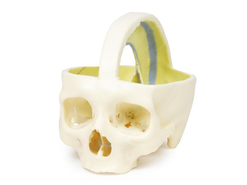
Skull and cranial cavity with the dural folds preserved.
Price on request
Available in Spain, Portugal and Andorra · delivery time given in the proposal
Downloadable data sheet — features, specifications and delivery time, in PDF.
Need a technical report? Ask us for it and we'll send it within 24 h →
Description
What this equipment is and what it's designed for.
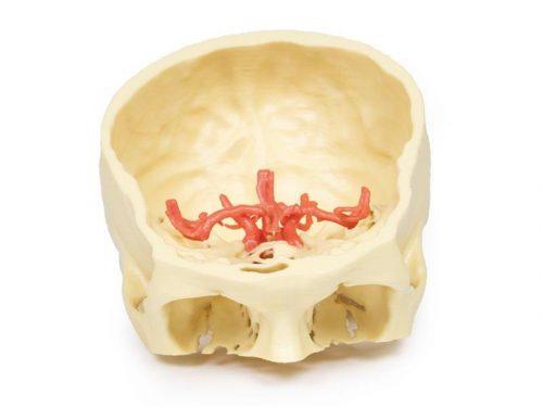
This high-quality 3D print of the skull and cranial cavity preserves key anatomical structures, including the dural folds. A thin strip of the calvaria has been retained in the mid-sagittal plane to maintain the attachment of the falx cerebri.
Preserved Tentorium Cerebelli
The tentorium cerebelli is carefully preserved in this model, clearly demonstrating the tentorial notch, which normally houses the mid-brain.
Highlighted Sella Turcica and Internal Carotid Arteries
In the region of the sella turcica, the entry points of the internal carotid arteries through the roof of the cavernous sinuses are highlighted in red, providing an intuitive visual guide for students and professionals.
Focus on Dural Venous Sinuses
The main purpose of this model is to showcase the disposition of the dural folds and the position of the dural venous sinuses, which are highlighted in pale blue. The following sinuses are clearly indicated:
– Superior sagittal sinus
– Inferior sagittal sinus
– Straight sinus
– Transverse sinuses
– Superior and inferior petrosal sinuses
– Sphenoparietal sinuses
– Cavernous sinuses
What you learn
Anatomical model modality. The competencies this equipment trains, in the language of the training plan.
- Identify the dura mater, the falx cerebri and the tentorium cerebelli.
- Follow the course of the dural venous sinuses.
- Relate the dural compartments to the routes of cerebral herniation.
Cases and pathologies
The clinical scenarios that can be simulated with this equipment.
- Extradural and subdural haematoma.
- Venous sinus thrombosis.
- Transtentorial herniation and its signs.
- Head injury with raised intracranial pressure.
Features and what's included
What the equipment does and what comes in the box, as described by the manufacturer.
Features
- 3D printed replica made from radiological data or from a dissected cadaveric specimen, not a stylised model: this is real anatomy.
- Developed and checked by the anatomists of the Human Anatomy Teaching Centre at Monash University.
- Colourised to distinguish muscle, nerve, artery, vein and bone, and to separate territories or segments.
- Not human tissue: no restrictions on transport, import or use in institutions without permission to work with cadavers.
- Avoids the ethical and preservation problems that come with plastination.
- Supplied with a detailed description of the anatomy represented in the specimen.
Services for this equipment
Services matter: the equipment gets commissioned, the staff know how to use it and maintenance is settled before you start. Include them in your budget and avoid having to raise a second purchase.
- Preventive maintenance We'll let you know when it's due
Documentation
You can download it now or we'll send it on request.
Other related equipment
They share a specialty with this one. They train different things: none of them replaces it.
Is this equipment going into a new centre?
If you're equipping a classroom or a whole centre, starting from the shopping list works out expensive. Start from the needs assessment.