

Reference 02678 · Anatomical model
3D ANATOMY - Deep dissection of the posterior body wall/ventral
- Anatomical model
- Anatomy
- Main product
- Medicine
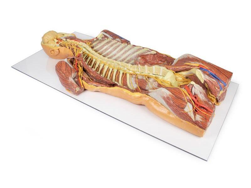
Ventral view of head, neck, thorax, abdomen and proximal thighs, with the central nervous system, the plexuses and the major vessels.
Price on request
Available in Spain, Portugal and Andorra · delivery time given in the proposal
Downloadable data sheet — features, specifications and delivery time, in PDF.
Need a technical report? Ask us for it and we'll send it within 24 h →
Description
What this equipment is and what it's designed for.
This 3D printed model provides a detailed ventral view of the head, neck, thorax, abdomen, and proximal thighs, offering insight into the central nervous system, nerve plexuses, and major vascular structures.
It serves as a complementary piece to the dorsal dissection model (MP1400).
Brain and Upper Spine
The facial skeleton is removed, exposing both cerebral hemispheres, parts of the Circle of Willis, and key arteries (vertebral, basilar, AICA, labyrinthine). Several cranial nerves (II, III, V, VI) and the right internal carotid artery are preserved. Below, the anterior spinal cord, vertebral arteries, and cervical and brachial plexus roots are visible, along with CN X, CN XII, and the sympathetic trunks.
Axillae and Upper Limbs
The anterior thoracic wall, clavicles, and first ribs are removed to expose both axillae, with clear views of the brachial plexus, axillary arteries, and surrounding musculature. The proximal upper limbs are preserved to mid-arm level.
Thorax and Lumbar Region
The spinal cord extends to the conus medullaris, with ventral nerve roots and sympathetic chains visible throughout. Splanchnic nerves, intercostal nerves, and abdominal wall nerves (e.g., ilioinguinal, genitofemoral, femoral) are preserved. On the right, removal of the psoas muscle exposes the lumbar plexus.
Pelvis and Proximal Thigh
The pelvic viscera are removed, but the pelvic floor, rectum, and lumbosacral plexus remain. Obturator nerves are visible entering the obturator canals, and the femoral vessels are retained in both femoral triangles.
This model is ideal for studying ventral CNS access, neurovascular pathways, and regional anatomical relationships across the entire torso.
What you learn
Anatomical model modality. The competencies this equipment trains, in the language of the training plan.
- Identify the deep muscle planes of the trunk wall.
- Follow the neurovascular bundles between the planes.
- Relate the dissection to the surgical approaches to the wall.
Cases and pathologies
The clinical scenarios that can be simulated with this equipment.
- Abdominal wall hernias.
- Thoracic and abdominal wall pain.
- Surgical approaches plane by plane.
- Teaching topographical anatomy of the trunk.
Features and what's included
What the equipment does and what comes in the box, as described by the manufacturer.
Features
- 3D printed replica made from radiological data or from a dissected cadaveric specimen, not a stylised model: this is real anatomy.
- Developed and checked by the anatomists of the Human Anatomy Teaching Centre at Monash University.
- Colourised to distinguish muscle, nerve, artery, vein and bone, and to separate territories or segments.
- Not human tissue: no restrictions on transport, import or use in institutions without permission to work with cadavers.
- Avoids the ethical and preservation problems that come with plastination.
- Supplied with a detailed description of the anatomy represented in the specimen.
Services for this equipment
Services matter: the equipment gets commissioned, the staff know how to use it and maintenance is settled before you start. Include them in your budget and avoid having to raise a second purchase.
- Preventive maintenance We'll let you know when it's due
Documentation
You can download it now or we'll send it on request.
Other related equipment
They share a specialty with this one. They train different things: none of them replaces it.
Is this equipment going into a new centre?
If you're equipping a classroom or a whole centre, starting from the shopping list works out expensive. Start from the needs assessment.