

Reference 11132 · Anatomical model
Functional and regional brain model, life-size, 5 parts - EZ Augmented Anatomy
- Anatomical model
- Anatomy
- Main product
- Medicine
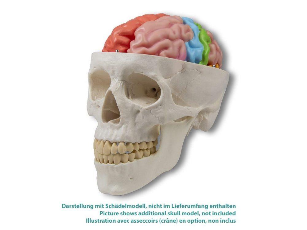
This life-size brain model shows the major anatomical landmarks and cortical areas of the brain in carefully painted color. The right half is painted to show the four lobes of the brain: parietal, temporal, occipital, and frontal. The left half comes apart in four pieces- the frontal and parietal, the temporal and occi
Price on request
Available in Spain, Portugal and Andorra · delivery time given in the proposal
Downloadable data sheet — features, specifications and delivery time, in PDF.
Need a technical report? Ask us for it and we'll send it within 24 h →
Description
What this equipment is and what it's designed for.
This life-size brain model shows the main anatomical landmarks and the cortical areas of the brain, carefully painted. The right half is painted to show the four cerebral lobes: parietal, temporal, occipital and frontal. The left half separates into four pieces: the frontal and the parietal, the temporal and the occipital, the brainstem and the cerebellum. It is painted to show 5 cortical areas: the primary motor cortex, the sensory cortex, the visual cortex, Broca’s area (motor or speech area), the auditory cortex and Wernicke’s speech area. This model fits our skull models ref. 4500, 4505, 4508, 4509, 4512, 4514, 4701 and 4708, and is compatible with the falx cerebri model ref. C925.
Augmented Anatomy App
The Augmented Anatomy app, together with this anatomical model, makes learning easier.
The app automatically recognises our anatomical models and displays the nomenclature in augmented reality. You can use it free of charge and with no time limit.
– Augmented reality learning app
– Free of charge and with no registration
– Nomenclature available anytime, anywhere
– Further links in the learning lexicon
Our augmented anatomy app works on the most common smartphones and tablets. Download the app
What you learn
Anatomical model modality. The competencies this equipment trains, in the language of the training plan.
- Distinguir los cuatro lóbulos cerebrales sobre la mitad derecha.
- Identificar las áreas corticales: motora primaria, sensitiva, visual, auditiva y las áreas de Broca y de Wernicke.
- Desmontar la mitad izquierda en sus cuatro piezas.
- Reconocer el tronco del encéfalo y el cerebelo por separado.
- Relacionar área cortical y función neurológica.
Cases and pathologies
The clinical scenarios that can be simulated with this equipment.
- Afasia de Broca y de Wernicke.
- Déficits motores y sensitivos por localización cortical.
- Alteraciones visuales de origen cortical.
- Docencia de neuroanatomía funcional.
Features and what's included
What the equipment does and what comes in the box, as described by the manufacturer.
Features
- Modelo de cerebro a tamaño natural en 5 partes.
- Mitad derecha pintada por lóbulos: parietal, temporal, occipital y frontal.
- Mitad izquierda separable en cuatro piezas: frontal y parietal, temporal y occipital, tronco del encéfalo y cerebelo.
- Mitad izquierda pintada por áreas corticales.
- Encaja en los cráneos ref. 4500, 4505, 4508, 4509, 4512, 4514, 4701 y 4708 y es compatible con la hoz del cerebro ref. C925.
Services for this equipment
Services matter: the equipment gets commissioned, the staff know how to use it and maintenance is settled before you start. Include them in your budget and avoid having to raise a second purchase.
- Preventive maintenance We'll let you know when it's due
Documentation
You can download it now or we'll send it on request.
Other related equipment
They share a specialty with this one. They train different things: none of them replaces it.
Is this equipment going into a new centre?
If you're equipping a classroom or a whole centre, starting from the shopping list works out expensive. Start from the needs assessment.