

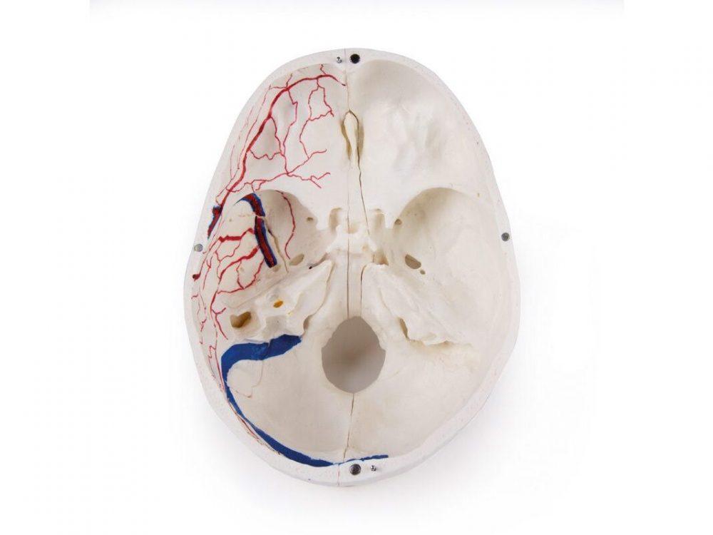
Reference 10991 · Anatomical model
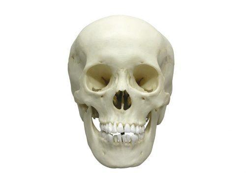
Deluxe demonstration skull; 14-part; for advanced studies
- Anatomical model
- Anatomy
- Main product
- Medicine
This skull model is an actual cast of a real human specimen and shows all anatomical structures in highest detail. It is made for students in anatomy, medicine, surgery, otolaryngology, ophthalmology and dentistry. The Skull is intricately sectioned and reassembled with metal and magnet connections. The calvarium is s
Price on request
Available in Spain, Portugal and Andorra · delivery time given in the proposal
Downloadable data sheet — features, specifications and delivery time, in PDF.
Need a technical report? Ask us for it and we'll send it within 24 h →
Description
What this equipment is and what it's designed for.
This skull model is a genuine cast of a human specimen and shows all the anatomical structures in great detail. It is intended for students of anatomy, medicine, surgery, ENT, ophthalmology and dentistry. The skull is precisely sectioned and reassembled by means of metal and magnetic connections.
The cranial vault is sectioned horizontally, leaving the temporal bone and its sutures intact. The bony impressions of the superior sagittal sinus, the transverse sinus and the sigmoid sinus have been painted, as have the meningeal vessels. The base of the skull is sectioned sagittally so that one cut passes through one cribriform plate of the ethmoid and another cut, in the same plane, passes through the cribriform plate on the opposite side, leaving the crista galli and the perpendicular plate of the ethmoid intact, as well as the whole nasal septum. The structures of the anterior, middle and posterior cranial fossae are easily accessible. You can look directly into the nasal cavity, the turbinates, the nasal septum and the bony pharyngeal and nasopharyngeal spaces. The nasal septum separates from the bones surrounding it. The frontal sinuses are dissected on one side to show the whole sinus and, on the other, cut back to give full access to their interior. The relationship of this sinus with the nasal cavity is clearly appreciated and is especially useful for ENT specialists.
On one side of the skull the temporal bone has been left in situ. The other temporal bone is removable. Part of the mastoid and of the squama can be removed together with the tympanic antrum, exposing the inner ear. The three semicircular canals can be seen, as can the course of the facial nerve, which runs backwards and then downwards until it exits through the stylomastoid foramen. The removable temporal bone keeps the external auditory canal intact. An almost vertical section has been made through the squama and the mastoid process, extended inwards along the petrosquamous junction; on separating it, the position of the tympanic membrane can be appreciated. The carotid canal and the cochlea have been opened, exposing the internal auditory canal, and the course of the facial nerve has been represented. The oval window, the semicircular canals and the aditus ad antrum are visible.
The maxilla and the mandible expose the structures of the dentition: the roots, the bony margin of the alveolar process and the dental vessels and nerves. The maxillary sinus is opened by removing a bone flap. The teeth of the right hemimandible are sectioned to show the internal structure of the tooth.
What you learn
Anatomical model modality. The competencies this equipment trains, in the language of the training plan.
- Identificar las estructuras de las fosas craneales anterior, media y posterior.
- Reconocer el contenido del hueso temporal: conductos semicirculares, cóclea y trayecto del nervio facial.
- Localizar los senos frontales y maxilares y su relación con la cavidad nasal.
- Describir las impresiones de los senos venosos durales y de los vasos meníngeos.
- Explicar la estructura interna del diente y del proceso alveolar.
Cases and pathologies
The clinical scenarios that can be simulated with this equipment.
- Docencia de anatomía para medicina, cirugía y odontología.
- Formación en otorrinolaringología: oído medio e interno, senos paranasales.
- Estudio de las vías de abordaje quirúrgico de la base del cráneo.
- Clases de oftalmología centradas en la órbita ósea.
Features and what's included
What the equipment does and what comes in the box, as described by the manufacturer.
Features
- Vaciado real de una pieza humana, con 14 partes.
- Secciones precisas que se vuelven a montar con uniones metálicas y magnéticas.
- Bóveda craneal seccionada en horizontal, con el hueso temporal y sus suturas intactos.
- Un hueso temporal extraíble, con el oído interno accesible y el conducto auditivo externo íntegro.
- Impresiones óseas de los senos sagital superior, transverso y sigmoideo pintadas.
- Maxilar y mandíbula con raíces, vasos y nervios dentales a la vista; seno maxilar abrible.
Services for this equipment
Services matter: the equipment gets commissioned, the staff know how to use it and maintenance is settled before you start. Include them in your budget and avoid having to raise a second purchase.
- Preventive maintenance We'll let you know when it's due
Documentation
You can download it now or we'll send it on request.
Other related equipment
They share a specialty with this one. They train different things: none of them replaces it.
Is this equipment going into a new centre?
If you're equipping a classroom or a whole centre, starting from the shopping list works out expensive. Start from the needs assessment.