

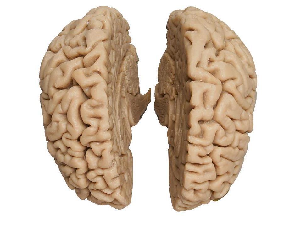
Reference 02667 · Anatomical model
3D ANATOMY - 3D human brain
- Anatomical model
- Nervous system
- Main product
- Medicine
Human brain moulded and cast from an actual specimen, in two separable hemispheres.
Price on request
Available in Spain, Portugal and Andorra · delivery time given in the proposal
Downloadable data sheet — features, specifications and delivery time, in PDF.
Need a technical report? Ask us for it and we'll send it within 24 h →
Description
What this equipment is and what it's designed for.
This Human Brain is molded and cast from an actual specimen. Custom-formulated resin and the production process permit us to represent a level of accuracy and detail necessary for anatomy instruction. The left and right hemispheres are separated, allowing for an interior view of the corpus callosum and cingulate gyrus, as well as the hypothalamus, pons, and cerebellum. Two different models available, each fits in a specific skull. Skulls not included.
Fits into our male skull model 4710.
What you learn
Anatomical model modality. The competencies this equipment trains, in the language of the training plan.
- Identify lobes, sulci and fissures on the cerebral surface.
- Locate the basal ganglia, the ventricular system, the brainstem and the cerebellum.
- Relate each area to its function and to the deficit caused by a lesion there.
- Orient the model against the sections seen on CT and MRI.
Cases and pathologies
The clinical scenarios that can be simulated with this equipment.
- Stroke by affected arterial territory.
- Brain tumours and mass effect.
- Epilepsy and localisation of the focus.
- Hydrocephalus and cerebrospinal fluid circulation.
Features and what's included
What the equipment does and what comes in the box, as described by the manufacturer.
Features
- 3D printed replica made from radiological data or from a dissected cadaveric specimen, not a stylised model: this is real anatomy.
- Developed and checked by the anatomists of the Human Anatomy Teaching Centre at Monash University.
- Colourised to distinguish muscle, nerve, artery, vein and bone, and to separate territories or segments.
- Not human tissue: no restrictions on transport, import or use in institutions without permission to work with cadavers.
- Avoids the ethical and preservation problems that come with plastination.
- Supplied with a detailed description of the anatomy represented in the specimen.
Services for this equipment
Services matter: the equipment gets commissioned, the staff know how to use it and maintenance is settled before you start. Include them in your budget and avoid having to raise a second purchase.
- Preventive maintenance We'll let you know when it's due
Documentation
You can download it now or we'll send it on request.
Other related equipment
They share a specialty with this one. They train different things: none of them replaces it.
-
 3D ANATOMY - Cubital fossa (muscles, major nerves and brachial artery)
See details →
-
 3D ANATOMY - Left shoulder, deep dissection of the joint, musculature, nerves and vessels
See details →
-
 3D ANATOMY - Nervous system dissection
See details →
-
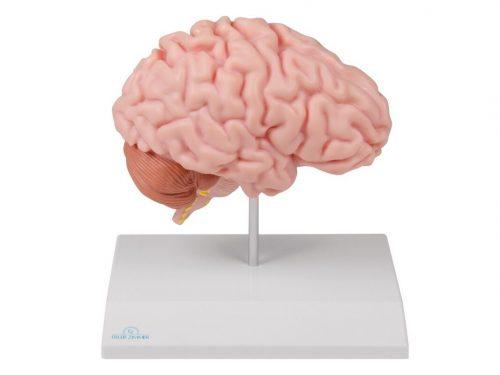 Anatomical brain half, life-size - EZ Augmented Anatomy
See details →
-
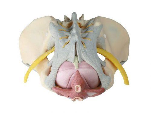 Anatomical model of female pelvis with ligaments, nerves and musculature
See details →
-
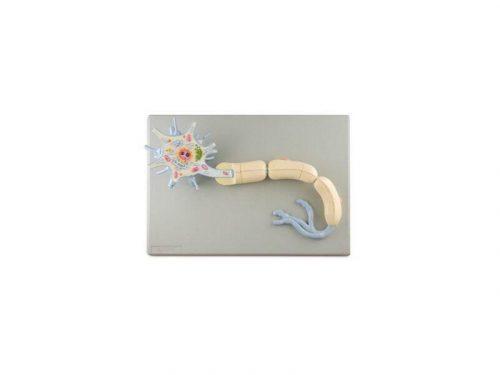 Anatomical model of neuron, Magnification x2500 - 2 Parts
See details →
Is this equipment going into a new centre?
If you're equipping a classroom or a whole centre, starting from the shopping list works out expensive. Start from the needs assessment.