

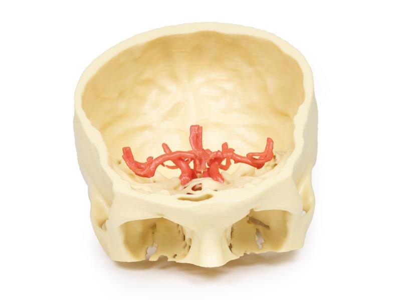
Reference 02672 · Anatomical model
3D ANATOMY - Circle of Willis
- Anatomical model
- Anatomy
- Main product
- Medicine
Intracranial arteries supplying the brain, reconstructed from segmented angiographic data.
Price on request
Available in Spain, Portugal and Andorra · delivery time given in the proposal
Downloadable data sheet — features, specifications and delivery time, in PDF.
Need a technical report? Ask us for it and we'll send it within 24 h →
Description
What this equipment is and what it's designed for.
This highly detailed 3D printed model offers a precise representation of the intracranial arteries supplying the human brain, created from segmented angiographic imaging data. The model displays vascular structures that cannot be physically dissected and viewed in situ, making it a unique and valuable teaching tool.
Vertebral and Basilar Arteries
The paired vertebral arteries are shown entering the cranial cavity through the foramen magnum, where they unite to form the basilar artery. The basilar artery travels along the brainstem and terminates by branching into the posterior cerebral arteries. Just before this bifurcation, the superior cerebellar arteries arise, supplying parts of the cerebellum and midbrain.
Internal Carotid Arteries and Carotid Siphon
The internal carotid arteries (ICAs) are traced from their entry at the carotid canal within the temporal bone. From there, they course medially and anteriorly through the petrous portion of the bone and emerge via the upper opening of the foramen lacerum. While the cavernous sinus is not shown, the model beautifully captures the characteristic S-shaped carotid siphon on both sides, clearly positioned lateral to the sella turcica and passing medial to the anterior clinoid processes.
Major Cerebral Arteries and the Circle of Willis
Each internal carotid artery divides into the anterior cerebral artery (ACA) and middle cerebral artery (MCA). The posterior communicating arteries, connecting the posterior cerebral arteries (PCAs) to the MCAs, are distinctly visible, allowing for an excellent demonstration of the posterior portion of the Circle of Willis.
The anterior communicating artery, completing the circle between the paired anterior cerebral arteries, is also present—though its visibility is limited due to the close proximity of the ACAs.
What you learn
Anatomical model modality. The competencies this equipment trains, in the language of the training plan.
- Recognise the arteries that form the circle of Willis and their main branches.
- Relate each artery to the cerebral territory it supplies.
- Identify the most frequent anatomical variants of the circle.
- Place the usual sites of aneurysm formation.
Cases and pathologies
The clinical scenarios that can be simulated with this equipment.
- Cerebral aneurysm and subarachnoid haemorrhage.
- Ischaemic stroke by arterial territory.
- Collateral circulation in carotid occlusion.
- Variants of the circle with clinical consequences.
Features and what's included
What the equipment does and what comes in the box, as described by the manufacturer.
Features
- 3D printed replica made from radiological data or from a dissected cadaveric specimen, not a stylised model: this is real anatomy.
- Developed and checked by the anatomists of the Human Anatomy Teaching Centre at Monash University.
- Colourised to distinguish muscle, nerve, artery, vein and bone, and to separate territories or segments.
- Not human tissue: no restrictions on transport, import or use in institutions without permission to work with cadavers.
- Avoids the ethical and preservation problems that come with plastination.
- Supplied with a detailed description of the anatomy represented in the specimen.
Services for this equipment
Services matter: the equipment gets commissioned, the staff know how to use it and maintenance is settled before you start. Include them in your budget and avoid having to raise a second purchase.
- Preventive maintenance We'll let you know when it's due
Documentation
You can download it now or we'll send it on request.
Other related equipment
They share a specialty with this one. They train different things: none of them replaces it.
Is this equipment going into a new centre?
If you're equipping a classroom or a whole centre, starting from the shopping list works out expensive. Start from the needs assessment.