

Reference 01824
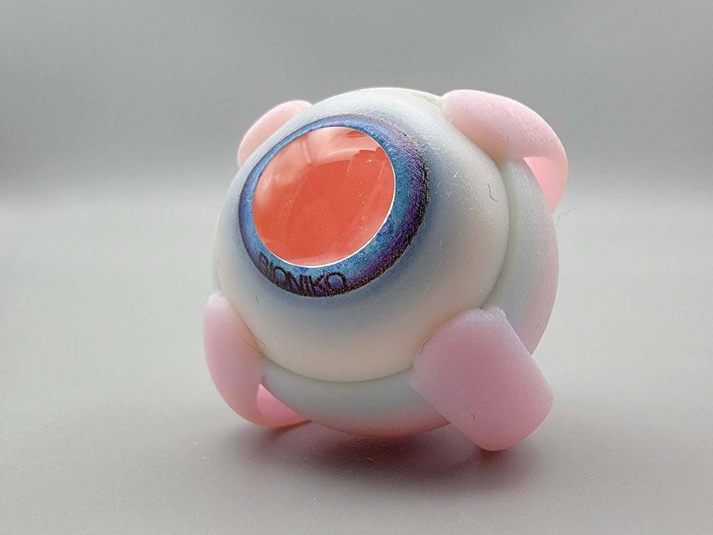
FUNDUS ADV - Advanced globe model with retinal image and optical element (1 pc)
- Ophthalmology
- Consumables
Eye simulator with retinal image, detachable colored iris anterior segment with flexible pars plana and optical element
Price on request
Available in Spain, Portugal and Andorra · delivery time given in the proposal
Downloadable data sheet — features, specifications and delivery time, in PDF.
Need a technical report? Ask us for it and we'll send it within 24 h →
Description
What this equipment is and what it's designed for.
Globe model with retinal image, detachable colored iris anterior segment with flexible pars plana and optical element. Includes assembly tool.Optimized for contact and non-contact systems.
Compatible with ROMANO or FLEX-ORBIT
What you learn
The competencies this equipment trains, in the language of the training plan.
- Vitrectomy
- ILM peeling
- Tractional retinal detachment (TRD)
- Fluid-air exchange (FAX)
- Laser procedures
- Foreign body
- Instrument manoeuvring skills
Related procedures: Retina
Cases and pathologies
The clinical scenarios that can be simulated with this equipment.
- Argon laser
- Posterior vitrectomy
- Epiretinal membrane (ERM)
- Advanced scenarios (retinal detachment, vitreous haemorrhage)
Features and what's included
What the equipment does and what comes in the box, as described by the manufacturer.
Features
KEY FEATURES:- Photo-realistic fundus with real-scale macula, optic nerve and arcades.
- Removable optical element simulates refractive power (50D)
- Replaceable pars-plana can be used with real surgical sharps
- Trans-illuminate for easy visualization in a training setting
- Remove all screws from FLEX-ORBIT.
- Squeeze the orbit in the nasal-temporal plane to round the orbit cavity
- Insert the FUNDUS model with the OPTIC NERVE (9) on the nasal side, and the muscles properly oriented (inferior, superior, lateral, medial).
- Release the FLEX-ORBIT so the cavity returns to its original shape. The eye should be lightly held by the superior and inferior muscles.
- Fix FLEX-ORBIT in place by pressing downward on a smooth surface to engage the suction-cup. NOTE: Lift the suction release tab to remove FLEX-ORBIT from surface. DO NOT PULL ON THE ORBIT!
- DISASSEMBLY: Gently push the optical element (1) (if needed) into the eye. Insert the legs of the assembly tool through the iris aperture and leverage the anterior segment out.
- ASSEMBLY: Slide the anterior segment support ring (4) under three muscle insertions (6) and use the assembly tool tip to help it under the fourth muscle insertion. Press on all quadrants to make sure the support ring is properly seated and no gaps are visible.
- Fill with vitreous substitute of choice such as egg white. Fillseal channel (5) with dispersiveviscoelastic before assembling to create a better fluid seal.
Includes
- 1 FUNDUS globe model with retinal image
- 1 FUNDUS anterior segment
- 1 Optical element
- 1 Storage box
Services for this equipment
Services matter: the equipment gets commissioned, the staff know how to use it and maintenance is settled before you start. Include them in your budget and avoid having to raise a second purchase.
- Preventive maintenance We'll let you know when it's due
Documentation
You can download it now or we'll send it on request.
Other related equipment
They train the same technique. Each one approaches it in its own way: none of them replaces this one.
-
 Digital eye examination trainer
See details →
- FUNDUS - Advanced retina scenarios kit See details →
-
 FUNDUS ADVANCED KIT
See details →
-
 FUNDUS BULLSEYE - Globe model with graduated reticle for field-of-view demonstration (1 pc)
See details →
-
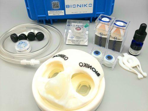 FUNDUS ROMANO - Retina surgery starter kit with intrascleral haptic fixation
See details →
- OphthoSim Mobile - 1 Student See details →
Is this equipment going into a new centre?
If you're equipping a classroom or a whole centre, starting from the shopping list works out expensive. Start from the needs assessment.