

Reference 02675 · Anatomical model
3D ANATOMY - Paranasal sinus model
- Anatomical model
- Anatomy
- Main product
- Medicine
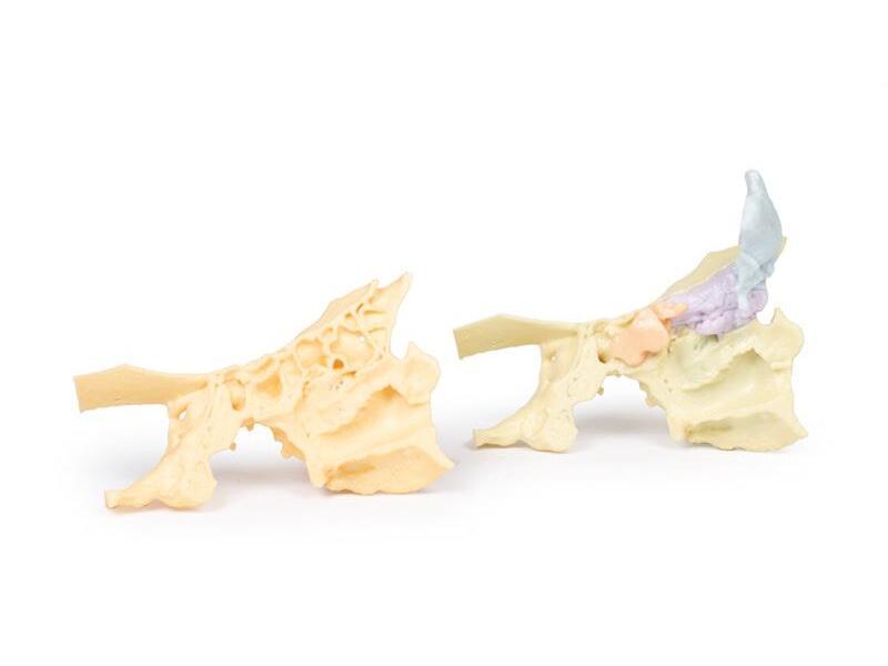
Paranasal sinuses obtained by segmenting the internal spaces from CT imaging.
Price on request
Available in Spain, Portugal and Andorra · delivery time given in the proposal
Downloadable data sheet — features, specifications and delivery time, in PDF.
Need a technical report? Ask us for it and we'll send it within 24 h →
Description
What this equipment is and what it's designed for.
This unique 3D model is derived from dural skull (Print #8) data, obtained via CT imaging and segmentation of internal spaces. Parts of the skull have been retained, while sections or windows have been removed to expose the paranasal sinuses.
Frontal Sinuses
The paired frontal sinuses are highlighted in blue. The right frontal sinus is fully exposed, showing the frontonasal ostium, which drains as a funnel-shaped tube into the infundibulum of the middle meatus of the nasal cavity. The left frontal sinus remains partially surrounded by frontal bone, illustrating the natural anatomy.
Ethmoid and Maxillary Sinuses
The ethmoid sinuses (purple) are visible only on the left side, with the medial wall of the orbit preserved, formed by the orbital plate of the ethmoid bone. The maxillary sinus (green) on the left is partially exposed and partially retained within the maxilla, demonstrating its close relationship to the inferior orbit. The opening of the maxillary sinus into the lateral wall of the nose is subtly indicated in the middle meatus.
Sphenoid Sinus
The left sphenoid sinus (pink) is embedded within the sphenoid bone, which has been partly digitally removed. Despite this, the impression for the internal carotid artery remains clearly identifiable, highlighting critical anatomical relationships.
What you learn
Anatomical model modality. The competencies this equipment trains, in the language of the training plan.
- Identify the paranasal sinuses and their drainage pathways.
- Recognise the landmarks of the middle meatus and the ostiomeatal complex.
- Relate the anatomy to the steps of endoscopic sinus surgery.
- Place the neighbouring structures at risk: orbit and skull base.
Cases and pathologies
The clinical scenarios that can be simulated with this equipment.
- Acute and chronic sinusitis.
- Sinonasal polyposis.
- Orbital and intracranial complications of sinusitis.
- Planning endoscopic sinus surgery.
Features and what's included
What the equipment does and what comes in the box, as described by the manufacturer.
Features
- 3D printed replica made from radiological data or from a dissected cadaveric specimen, not a stylised model: this is real anatomy.
- Developed and checked by the anatomists of the Human Anatomy Teaching Centre at Monash University.
- Colourised to distinguish muscle, nerve, artery, vein and bone, and to separate territories or segments.
- Not human tissue: no restrictions on transport, import or use in institutions without permission to work with cadavers.
- Avoids the ethical and preservation problems that come with plastination.
- Supplied with a detailed description of the anatomy represented in the specimen.
Services for this equipment
Services matter: the equipment gets commissioned, the staff know how to use it and maintenance is settled before you start. Include them in your budget and avoid having to raise a second purchase.
- Preventive maintenance We'll let you know when it's due
Documentation
You can download it now or we'll send it on request.
Other related equipment
They share a specialty with this one. They train different things: none of them replaces it.
Is this equipment going into a new centre?
If you're equipping a classroom or a whole centre, starting from the shopping list works out expensive. Start from the needs assessment.