CARACTERÍSTICAS
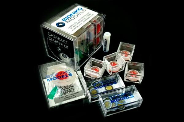
MAIN COMPONENTS OF THE PACKAGE:
AVH HOLDER:
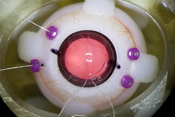
- The holder features a vitreouscavity that can be filled with vitreous substitute through a port using a standard syringe (5cc recommended). The soft seal mates with the OKULO anterior segment model when inserted in the holder, allowing for vitreous loss to be controlled via the syringewhen a rupture is made in the posterior capsule of a cataract lens or an empty bag lens.
- As with all BIONIKO holders, the AVH has flexible neck to simulate natural eye movement and is coupled to a suction cup for fixation to any flat surface.
KULO BR8:
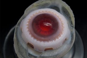
- The OKULO ANTERIOR SEGMENT, with BROWN iris and 8 mm pupil (BR8) features a self-sealing, transparent cornea, a pigmented angle, a flexible iris, sulcus space and a compliant sclera, allowing multiple surgical scenarios to be performed.
MEDIUM AND HARD CATARACT LENSES:
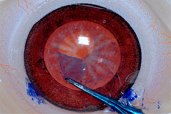
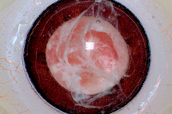
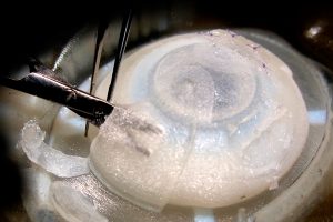
- Lenses for simulating Capsulorhexis, hydro-dissection and hard nucleus phacoemulsification scenarios.
LENS TOOL:
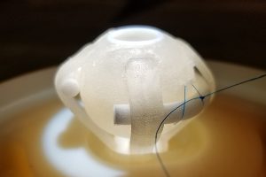
- Assembly tool for OKULO and RHEXIS lenses.
LENS ASSEMBLY:
There are a variety of lens options, each one suitable for different scenarios. Refer to the lens instructions for use for additional information.
- Surgical lenses allow capsulorhexis and phacoemulsification maneuvers to be practiced. The lens can be replaced to re-use the ANTERIOR SEGMENT multiple times.
- To insert, align the LENS with the CILIARY BODY, orienting the ANTERIOR CAPSULE (shiny side) facing the iris.
- Gently tuck the ATTACHMENT ZONULE into the CILIARY BODY GROOVE. Engage one zonule sector first and then press around the entire zonule to engage 360 degrees. Lubricating the lens or ciliary body with water may help engagement.
- Press zonule lightly: Lens should be in CB GROOVE and not in CB SULCUS. Hold ANTERIOR SEGMENT by the SUPPORT RING when inserting the lens. DO NOT PULL/PUSH on the cornea!
Descripción
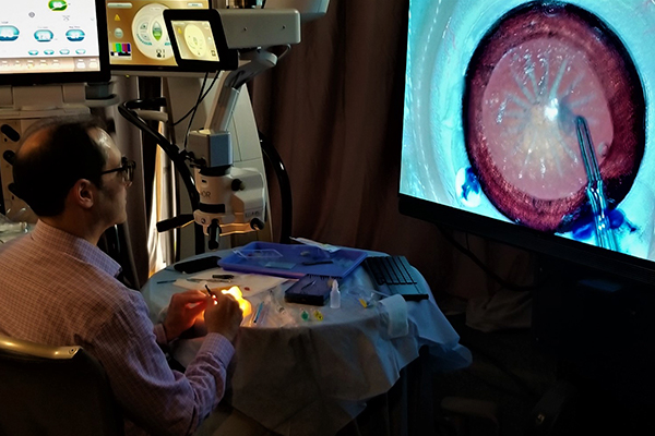
Este paquete de simulación es el producto básico perfecto para los usuarios que se inician en plataformas de simulación quirúrgica ocular y contiene en una sola caja todos los productos necesarios para tener un laboratorio experimental de cirugía de cataratas fácil de preparar.
Optimice su experiencia de formación de cataratas con una simulación realista de los principales pasos quirúrgicos: incisiones, relleno viscoelástico, capsulorrexis, hidrodisección, facoemulsificación e implantación de lentes intraoculares.

