SKILLS
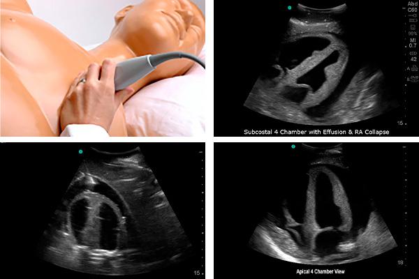
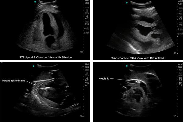
- Two-dimensional, three-dimensional and four-dimensional ultrasound scanning
- Ultrasound-guided pericardiocentesis
- Observation of pericardial effusion
- Site selection
- Optimal puncture route during the procedure
- Confirmation of optimal needle tip position
- Fluid removal