CASES AND PATHOLOGIES
SETUP WITH ANIMAL OR DONOR TISSUE:
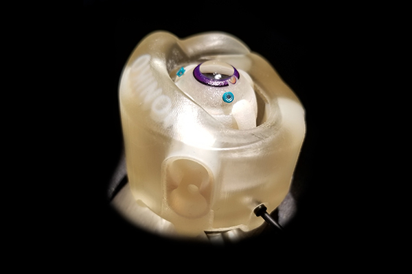
Socket adapter is not needed for use with animal ordonor tissue.
- Retract the four anterior screws.
- Line the Orbit cavity with a thin gauze pad. This acts as a fluid barrier and helps retain tissue.
- Position the globe such that the globe equator is at or below the anterior screws.
- Adjust posterior screws to adjust eye depth. Inserting these screws will support the eye higher in the orbit; retracting these screws will support the eye deeper in the orbit. All screws should be even for best results.
- Adjust the anterior screws to restrict anterior eye movement. All screws should be even for best grip.
- TIP: Needles can be inserted into or through the FLEX-ORBIT body for additional eye fixation.
- Increase intra-ocular pressure (IOP) if desired; adjust screws so that the anterior screws lie slightly above the equator of the eye. The external pressure of the screws against the sclera increases IOP.
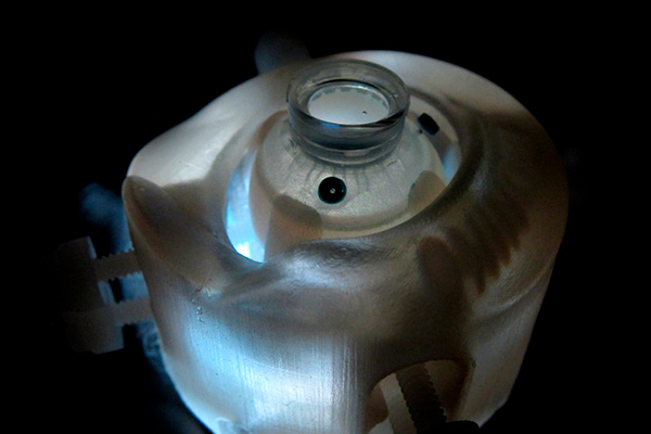
- Position FLEX-ORBIT according to the desired approach (temporal, superior).
- Fix the FLEX-ORBIT in place by pressing downward on a smooth surface to engage the suction-cup.
ADDITIONAL MODULES
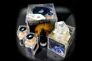
ACCESSORIES (sold separately):
There are several accessories available that can complement and enhance the simulation/training experience and fidelity of the FLEX-ORBIT platform:
- POSTERIOR-SEGMENT model: Allows attachment of anterior segment models, such as the RHEXIS and KERATO tasks, providing them with the realism and mobility of wholeglobe models.
- EYELID model: Attaches to the adapter groove (10) on the FLEX-ORBIT, adding the realism and restriction of eyelids.
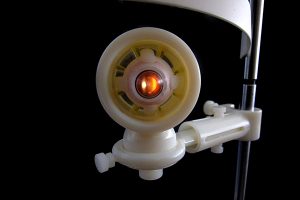
Description
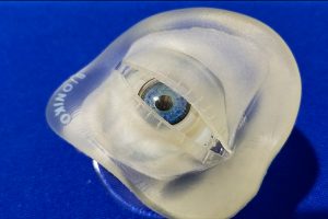
The FLEX-ORBIT is a universal eye-holder, compatible with BIONIKO synthetic models and ex-vivo biological tissue. It provides the user with a compact yet realistic frame of reference, including anatomical landmarks such as the brow and eye movement simulation that provide an added challenge.
This platform is ideal for use in the wet-lab, research lab, and tradeshow floor to communicate with colleagues, patients and clients.
KEY FEATURES:
- Portable and effective holder for anterior segment and whole globe models, including biological tissue.
- Adds anatomical constraints and eye motion to the simulation scenario.
Compatible with all models (except LinK)