CHARACTERISTICS
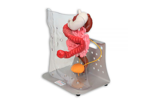
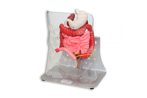
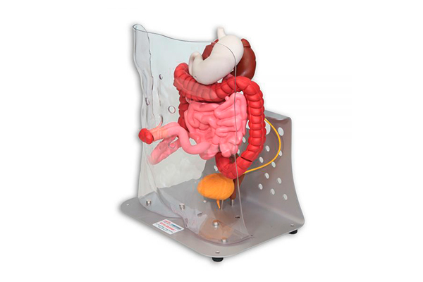
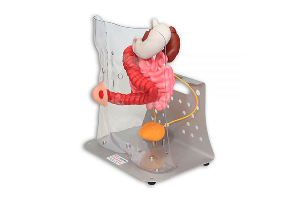
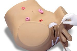
Otto Ostomy™ was modeled from a patient?s CT scan. The color coded organs displayed include:
- Stomach
- Small Intestine
- Large Intestine
- Rectum
- Kidneys
- Ureters
- Bladder
The small intestine, large intestine, rectum and bladder are all removable to aid in teaching procedures where these organs might be removed.
The ureters can be removed from the bladder and reinserted into the ileal conduit to show how a urostomy functions. The flexible small and large intestines can be separated and attached to the backside of the stoma in the torso shell to demonstrate either an end or loop stoma. The large intestine can be separated at four different locations: Ascending colon, Transverse colon, Descending colon and the Sigmoid colon.
Description
Otto Ostomy™ Advanced Model is supplied with 18 stomas. The more healthcare providers understand ostomies, the better they will be able to educate and encourage their patients about this life-changing event.
Seeing the 3D digestive and urinary tracts and visualizing the location and function of the various organs is essential to learning, especially in those cases where cognitive processes or language may be an obstacle.

