SKILLS
- Diagnosis and treatment of joint diseases
- Osteoarthritis
- Rheumatoid arthritis
- Puncture of the knee joint cavity
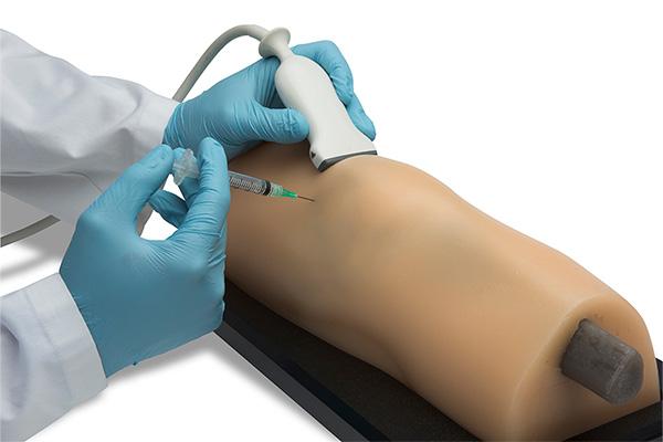
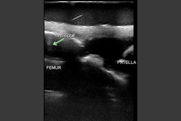
- Suction and injection of drugs under ultrasound guidance or according to body surface markers

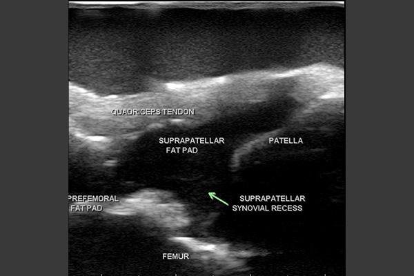
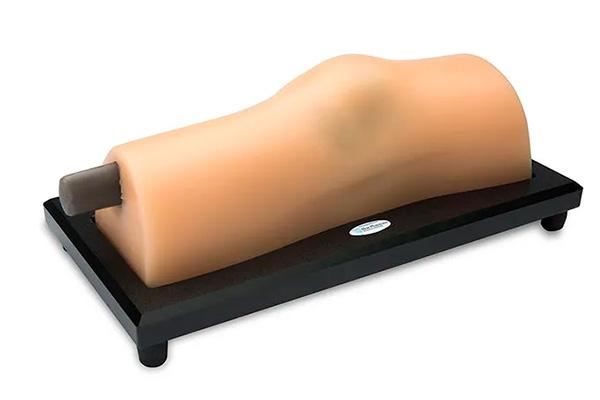
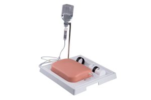
Knee phantom for musculoskeletal ultrasound and ultrasound-guided infiltration incorporates all the anatomy necessary to teach, learn and practice the skills associated with injections and joint aspirations of the knee.
This phantom reproduces the tendons and ligaments of the knee to provide a truly realistic training environment. Learn and practice the psychomotor skills associated with ultrasound-guided or blind insertion of knee injections and aspirations associated with osteoarthritis and rheumatoid arthritis.
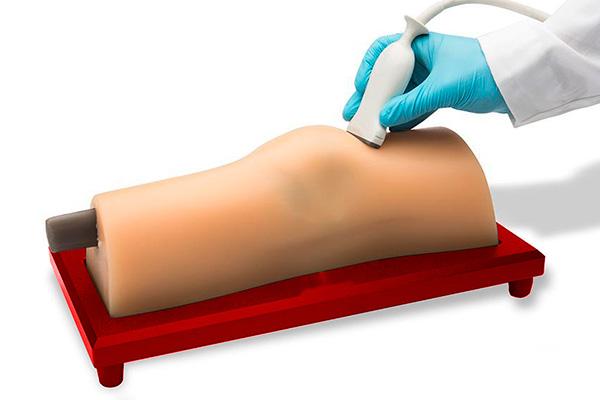
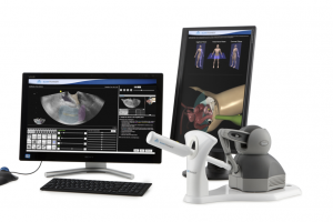
The compact system for medical professionals wishing to learn scanning and diagnostic skills in transvaginal and transabdominal ultrasound.
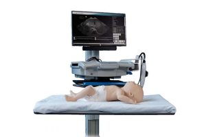
BabyWorks Sam is an ultra-realistic baby manikin offering a safe and effective training tool for Point of Care Ultrasound (PoCUS) and Echocardiography in pediatric and neonatal care.