

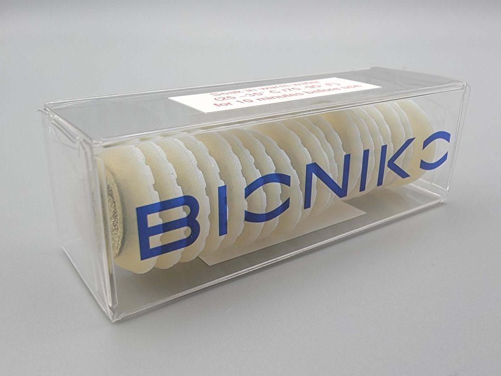
Reference 1756
CORDELIA - Corneal excision
- Emergencies
- Consumables
In situ corneal recovery and trauma suturing ocular surgery simulator. For use with BASIC ORBIT or EXOS HM/FLEX ORBIT HOLDERS.
Price on request
Available in Spain, Portugal and Andorra · delivery time given in the proposal
Downloadable data sheet — features, specifications and delivery time, in PDF.
Need a technical report? Ask us for it and we'll send it within 24 h →
Description
What this equipment is and what it's designed for.
CORDELIA focuses on providing a realistic anatomical reference for practicing gross corneal dissection techniques like in-situ corneal excision, and corneal trauma repair.
The model anatomy includes a scleral layer and choroid layer. These layers are separated posterior to the limbus, simulating the supra-choroidal space, and are attached at the spur. The user can practice detaching the tissue at the spur with minimal stress to the endothelium, an essential technique for corneal recovery.
What you learn
The competencies this equipment trains, in the language of the training plan.
- Corneal dissection techniques
- Corneal suturing techniques (trauma scenarios)
- In-situ corneal excisions
- Instrument manouvering skills
Cases and pathologies
The clinical scenarios that can be simulated with this equipment.
- Corneal trauma suturing
Features and what's included
What the equipment does and what comes in the box, as described by the manufacturer.
Features
FEATURES:- Cornea provides realistic feel and accurate anatomical proportions to practice corneal suturing, and the iris feature adds to the model's realism.
- The structural ring at the base of the model maintains the model's shape and the notches prevent unwanted rotation of the model during trephination.
- Any BIONIKO holder works for corneal suturing tasks. A Standard ORBIT Holder (sold separately) is recommended to practice in-situ corneal recovery.
- The ORBIT model serves as a base for the CORDELIA model and provides reference and realism by challenging the user to manipulate instruments according to the facial structures around the eye. There are both right (R) and left (L) ORBIT models to practice both approaches.
- Add water into the anterior chamber of the model with a dropper or under the tap.
- Soak in water (~37ºC for best results) for 10 minutes for cornea to hydrate.
- Load CORDELIA model in the ORBIT. The ORBIT has a flexible “eyelid” that receives and secures the CORDELIA model. Use a couple of waterdrops to moisten the eyelid and socket area. Insert the edge of the model under the superior eyelid and push the inferior sclera from the structural ring into the socket to load the model.
- Fix the ORBIT in place by pressing downward on a smooth surface to engage the suction-cup. Orient the ORBIT according to the desired approach: Superior (S), Nasal (N), Inferior (I) or Temporal (T).
- Hydrate/lubricate model during task by adding water. Model's compliance may decrease if dehydrated, so more is better than less.
- IN-SITU EXCISION: Practice corneal excision techniques, including steps of trephination, sclerotomy and corneal teasing (dettachment from spur).
- CORNEAL SUTURING: CORDELIA can also be used to practice corneal suturing techniques by creating a laceration or other trauma scenarios.
Includes
- 20 Corneal excision simulator
- 1 Storage box
Services for this equipment
Services matter: the equipment gets commissioned, the staff know how to use it and maintenance is settled before you start. Include them in your budget and avoid having to raise a second purchase.
- Preventive maintenance We'll let you know when it's due
Documentation
You can download it now or we'll send it on request.
Other related equipment
They train the same technique. Each one approaches it in its own way: none of them replaces this one.
Is this equipment going into a new centre?
If you're equipping a classroom or a whole centre, starting from the shopping list works out expensive. Start from the needs assessment.